Summary
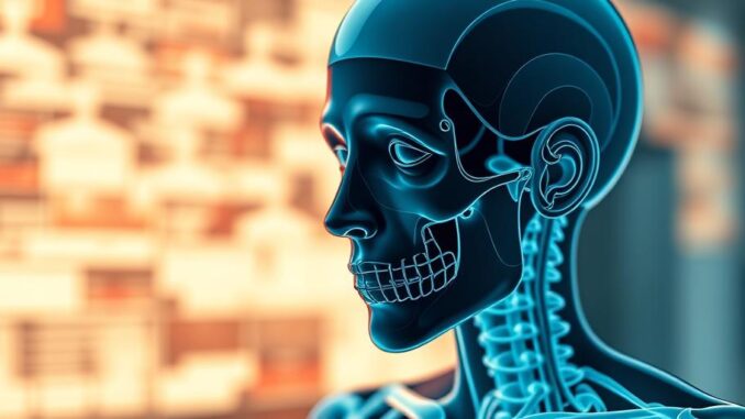
3D medical imaging is revolutionizing healthcare by providing detailed views of organs, enabling faster diagnoses and personalized treatments. This technology utilizes advanced software and imaging techniques like CT, MRI, and ultrasound to create interactive 3D models, which are transforming medical education, surgical planning, and disease monitoring. From virtual colonoscopies to tumor growth tracking, 3D organ visualization offers numerous benefits for both patients and healthcare professionals.
Secure patient data with ease. See how TrueNAS offers self-healing data protection.
** Main Story**
Medical technology is constantly pushing boundaries, and let me tell you, one of the coolest advancements I’ve seen lately is 3D organ visualization. It’s really taking medical imaging to a whole new level, leaving behind those old 2D X-rays for interactive, three-dimensional models. These detailed visualizations are incredibly beneficial, not just for us healthcare professionals, but more importantly for patients as well; it’s impacting everything from diagnoses and treatment plans to how we educate future doctors.
Crafting the 3D Masterpiece: A Symphony of Tech
So, how do they actually do it? Well, it all starts with some seriously impressive imaging techniques. Think CT scans, MRIs, and even good old ultrasound – these all provide the raw data, basically capturing detailed cross-sectional images. Then, specialized software steps in. And it’s not your average program, this stuff uses sophisticated algorithms to reconstruct the images into a 3D model you can rotate, zoom, and generally play around with.
Doctors can now examine an organ from every possible angle. It’s kind of mind-blowing, actually. Think about the potential there. I mean, it wasn’t that long ago that we were stuck with flat images, now this! All those improvements in automated image processing and 3D modeling? They’re really driving this field forward and leading to ever more sophisticated and accurate representations of what’s inside us.
3D in Action: Revolutionizing Medicine
Let’s talk about how this tech is really changing things.
-
Diagnosis and Treatment Planning
3D organ visualization is seriously changing the game for diagnosis and treatment. I saw firsthand how virtual colonoscopies use 3D models of the colon to detect polyps; you know, offering a less invasive alternative to the traditional methods. Then there’s cardiology. 3D imaging helps doctors assess heart function and spot any blood vessel abnormalities, which in turn helps them plan interventions like angioplasty or bypass surgery. And neurologists? They’re using 3D brain imaging to pinpoint tumors, which is obviously crucial for surgical planning.
This level of precision? It allows for much more targeted interventions, minimizing damage to the surrounding healthy tissue. The result? Much better outcomes for patients, it’s pretty cool. Think about it, a few years ago, a surgeon would have to go in almost blind; now they have a detailed, interactive map! A misplaced comma, here or there, doesn’t change that fact.
-
Medical Education and Training
This is a personal favorite. I remember struggling with anatomy textbooks back in med school. Now imagine learning anatomy and physiology with interactive 3D models – it’s a game changer. Surgical simulations using 3D models allow surgeons to practice complex procedures in a virtual environment. And that’s before they even think about touching a real patient! You can find 3D anatomical atlas apps on Google Play, which offer models and descriptions of various organ systems. It’s a hands-on, visual approach. It boosts understanding and skill, and that leads to better patient care, what’s not to like?
-
Monitoring Disease Progression
And don’t forget about disease monitoring. By comparing 3D images over time, doctors can track changes in organ structure and function. Oncologists, for instance, can use 3D imaging to monitor tumor growth. This helps them evaluate how well a patient is responding to chemotherapy. Or other cancer therapies. This longitudinal analysis is invaluable for making informed decisions about adjusting treatment plans. I mean, you want to catch these things early, right? It really maximizes a patient’s chances of success.
Looking Ahead: The Future is 3D
The field of 3D medical imaging is not slowing down, it’s accelerating. Researchers are developing new imaging techniques, and software tools. I’m talking improved image resolution, more realistic models, and wider applications. Have you heard of terahertz (THz) imaging? It’s offering non-invasive, high-resolution visualization of cochlear structures, and other delicate tissues. These innovations promise precise diagnoses and personalized treatments. I, for one, can’t wait to see what’s next.
-
High-Resolution Mapping of Enzyme Activity
New probe molecules are allowing scientists to create 3D images of enzyme activity in whole organs. This provides insights into biological processes at a microscopic level.
-
3D X-Ray Imaging
Recent milestones in 3D X-ray technology enable high-resolution CT scans of dense objects. You get detailed internal views without invasive procedures. Sounds good, right?
-
AI-Powered Diagnostic Software
Artificial intelligence algorithms are being integrated into 3D imaging software. They analyze medical data and predict the likelihood of certain medical conditions. This can lead to faster and more accurate diagnoses, so the computers are coming for our jobs, but, at least, they’ll tell us what’s wrong with us!
Ultimately, with its ability to transform complex medical data into visual representations, 3D organ visualization is modern medicine’s cornerstone. It’s paving the way for a future of personalized healthcare. And that, my friends, is something to get excited about.


The application of 3D visualization to medical education is compelling. Beyond surgical simulations, how might these models be used to enhance patient understanding of their conditions and treatment options, potentially improving adherence and overall satisfaction?
That’s a fantastic point! Visualizing their own anatomy could empower patients to actively participate in their care. Imagine interactive models showing how a treatment works or the impact of lifestyle choices. This could definitely boost understanding and commitment. Let’s explore ways to make this technology more accessible to patients!
Editor: MedTechNews.Uk
Thank you to our Sponsor Esdebe
Virtual colonoscopies sound amazing! Forget the prep, can we get a 3D rendering of the fridge before I decide on midnight snacks? Precision in diagnosis and snacking – that’s the future I want to live in!
That’s hilarious! A 3D fridge scan would definitely revolutionize snack decisions. Imagine an app that calculates the nutritional value of your potential midnight feast in real-time. Maybe we’re closer to that future than we think!
Editor: MedTechNews.Uk
Thank you to our Sponsor Esdebe
The advancements in surgical simulations using 3D models are particularly exciting. This technology not only refines surgical skills in a virtual environment, but also offers opportunities for collaborative training across geographical boundaries, potentially democratizing access to specialized surgical expertise.
Absolutely! The collaborative training aspect is huge. Imagine surgeons in different countries working together on a virtual case, sharing expertise in real-time. This could significantly improve surgical outcomes and standardize best practices globally. It’s an exciting prospect for the future of medical collaboration!
Editor: MedTechNews.Uk
Thank you to our Sponsor Esdebe