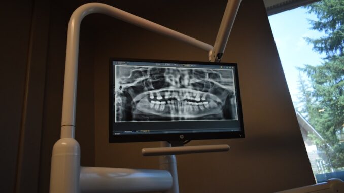
The New Frontier: Revolutionizing X-ray Imaging for Safer, Sharper Diagnostics
It’s incredible, isn’t it, how rapidly medical technology evolves? You’d think after more than a century, X-ray imaging, that stalwart of diagnostics, might’ve settled into a comfortable routine. But no, quite the opposite. We’re witnessing nothing short of a revolution in the field, one driven by an unrelenting pursuit of enhanced patient safety and unparalleled diagnostic precision. The core of this transformation, if you ask me, lies firmly in recent breakthroughs concerning detector technology, innovations that aren’t just incrementally improving image quality but are fundamentally recalibrating our understanding of radiation exposure and diagnostic capability.
From the moment Wilhelm Röntgen peered at the bones in his wife’s hand, X-rays have been indispensable. Yet, for decades, the technology, while life-saving, carried an inherent trade-off: the necessary dose of ionizing radiation. Today, that narrative is dramatically shifting. Researchers, engineers, and clinicians are collaboratively pushing boundaries, giving us tools that deliver clearer images with significantly less exposure, opening up entirely new avenues for patient care. It’s a really exciting time to be involved in healthcare technology, I think.
Secure patient data with ease. See how TrueNAS offers self-healing data protection.
Foldable Detectors: A Leap Towards Unprecedented Mobility and Safety
One of the most genuinely promising advancements making waves right now is the emergence of foldable X-ray detectors. This isn’t just about making things smaller; it’s about fundamentally rethinking how and where we perform diagnostic imaging. Picture this: a highly sensitive, high-resolution X-ray detector that can be literally folded up, tucked away, and then deployed on demand. Sounds like science fiction, right? Well, it isn’t.
Researchers, particularly those at the King Abdullah University of Science and Technology, have been instrumental in this development. They’ve engineered a detector that leverages the remarkable properties of methylammonium lead bromide perovskite crystals. Now, perovskites are a class of materials that have been gaining immense attention across various tech sectors, primarily because of their exceptional light-harvesting and charge-transport properties. For X-ray detection, their high atomic number means they’re incredibly efficient at stopping X-ray photons, and their unique crystal structure allows for highly efficient conversion of X-ray energy into an electrical signal, minimizing the wasteful energy loss seen in older technologies.
What makes this particular iteration so ingenious is its cascade configuration. Think of it like a carefully arranged series of tiny, highly efficient nets designed to catch every single X-ray photon with precision, and almost zero wasted motion. This design isn’t just a clever arrangement; it’s pivotal for effectively minimizing background noise, which, as you know, is the bane of any imaging system. By doing so, the detector’s sensitivity skyrockets. What this means in practical terms is that you can achieve high-quality imaging at significantly lower radiation doses. We’re talking about capturing intricate details – the kind of resolution that let researchers visualize the tiny internal wiring of a USB cable or the precise entry point of a metal needle piercing a raspberry, all while reducing the patient’s exposure. It’s truly impressive, the detail you can glean from such low doses.
The implications of this foldable nature extend far beyond just technical specs, though. It offers substantial advantages in dynamic clinical environments like point-of-care settings and, especially, emergency departments, where rapid and accurate imaging can literally be the difference between life and death. Their inherent portability and user-friendliness empower healthcare providers to conduct bedside evaluations swiftly. Imagine the scenario: a patient in critical condition, unable to be moved to a dedicated radiology suite. With a foldable detector, the imaging comes to them, reducing discomfort, minimizing movement-related risks, and slashing diagnostic turnaround times. This drastically improves patient outcomes and streamlines the entire clinical workflow, a real godsend for overstretched medical teams.
Moreover, this reduction in radiation exposure aligns perfectly with the burgeoning global emphasis on patient safety. We’re all increasingly aware of the cumulative risks associated with ionizing radiation, and tools that help us achieve diagnostic clarity while mitigating those risks are not just desirable; they’re becoming essential. It’s about ‘ALARA’ – As Low As Reasonably Achievable – in practice, not just in theory. While challenges remain in terms of mass production and long-term material stability, the path these foldable detectors are paving feels incredibly promising, don’t you think?
Photon-Counting Detectors: Unlocking Spectral Information for Deeper Insight
Alongside the innovations in portability, another truly groundbreaking development is the advent of photon-counting detectors. This technology isn’t just an evolution; it’s a paradigm shift in how X-ray data is acquired and processed. Traditional X-ray detectors, often called ‘energy-integrating’ detectors, work by summing up all the X-ray energy that hits them, much like a rain gauge collects water without distinguishing between small drizzles and heavy drops. You get a total amount, but you don’t know the individual characteristics.
Photon-counting detectors, conversely, operate on an entirely different principle. They register individual X-ray photons as they arrive and, crucially, measure the precise energy level of each one. Imagine counting every single raindrop and, as you do, also measuring its size. This ability to differentiate photons by their energy opens up a completely new dimension in imaging: spectral imaging. This means we’re no longer just looking at a grayscale shadow; we’re gaining detailed, quantitative information about tissue composition. We can, for example, differentiate between iodine and calcium, which appear similarly dense on conventional X-rays, providing unprecedented clarity for diagnosing subtle abnormalities that might easily be overlooked with older methods. It’s like moving from a black-and-white photograph to a full-color, chemically analyzed digital image.
The clinical significance of this can’t be overstated. The first clinically approved photon-counting computed tomography (CT) system, cleared by the FDA in September 2021, marked a pivotal moment. This wasn’t merely a technological curiosity; it was a validated tool ready to transform patient care. Think about it: a CT scanner that can not only show you where a lesion is but also tell you, with greater certainty, what it’s made of. That’s a game-changer for oncologists, cardiologists, and many others.
Photon-counting detectors boast several distinct advantages over their conventional energy-integrating counterparts. You’re looking at a significantly improved signal-to-noise ratio, meaning clearer images with less ‘fuzziness.’ There’s also a substantial reduction in the X-ray dose delivered to the patient, a benefit that impacts every single individual undergoing imaging. Furthermore, their enhanced spatial resolution allows for the visualization of finer anatomical details. These benefits, collectively, translate into more accurate diagnoses, earlier detection of disease, and ultimately, better patient management and care. For instance, studies examining photon-counting mammography have shown remarkable improvements in diagnostic performance, all while reducing radiation exposure by as much as 60% compared to conventional techniques. That’s a significant win-win, isn’t it? We’re talking about potentially catching cancers earlier with less risk to the patient, and that’s exactly what healthcare innovation should be about.
Artificial Intelligence: The Intelligent Co-Pilot in Diagnostic Imaging
Beyond the detectors themselves, the broader ecosystem of X-ray imaging has been profoundly impacted by the seamless integration of artificial intelligence. This isn’t about replacing the human element, far from it; it’s about providing radiologists and other clinicians with an incredibly intelligent co-pilot, enhancing their capabilities and streamlining their demanding workflows. AI algorithms are now assisting across a spectrum of tasks, from the initial image acquisition right through to the final diagnostic interpretation.
Let’s break down where AI is making the biggest splashes. We’re seeing AI being deployed for sophisticated image reconstruction, taking raw data from the detector and turning it into a clear, usable image much faster and often with fewer artifacts than traditional methods. It’s also become incredibly adept at noise reduction, distinguishing genuine signals from the inherent ‘static’ in the image, and correcting for various artifacts—think patient motion or metallic implants that can obscure vital anatomical details. Perhaps one of the most exciting applications is in automated analysis. AI systems, trained on vast datasets of medical images, can now autonomously detect and highlight potential abnormalities, whether they’re subtle lung nodules, hairline fractures, or early signs of osteoporosis. This ability to draw a radiologist’s eye to areas of concern acts like a second, tireless pair of eyes, ensuring nothing important gets missed in the sheer volume of images they review daily.
This integration delivers a multi-faceted benefit. Firstly, it demonstrably improves diagnostic accuracy. By reducing noise and highlighting anomalies, AI gives radiologists a clearer canvas and a valuable prompt, allowing them to focus their expert attention on critical findings. Secondly, it drastically streamlines workflows. Imagine a system that can pre-sort studies by urgency, automatically measure anatomical structures, or even draft initial reports. This allows for faster and more efficient image interpretation, meaning patients get their diagnoses quicker, and treatment plans can be initiated without delay. It’s a huge step forward in operational efficiency.
Moreover, AI plays a crucial role in mitigating human error and reducing variability in interpretations. Even the most experienced radiologist can experience fatigue, leading to slight inconsistencies. AI, however, maintains a consistent level of scrutiny, leading to more reliable and reproducible imaging results across the board. As AI technology continues its rapid evolution, its role in medical imaging is only set to expand, promising new frontiers in areas like early disease detection, predictive diagnostics, and truly personalized treatment planning, where therapy is tailored not just to a disease, but to an individual patient’s unique biological profile. The synergistic relationship between advanced detectors and intelligent algorithms truly represents the future of medical imaging.
Miniaturization and Portability: Bringing Imaging to the Patient’s Bedside
For a long time, the X-ray machine was this imposing, fixed apparatus, almost a monument in the radiology department. You had to bring the patient to it. But today’s healthcare landscape demands flexibility, speed, and patient-centric care. This is precisely why the demand for truly portable and compact X-ray detectors has seen such a meteoric rise, particularly in dynamic environments like point-of-care settings, mobile imaging units, and, crucially, busy emergency departments.
These portable systems aren’t just convenient; they offer unparalleled flexibility, allowing healthcare providers to bring imaging capabilities directly to the patient. This translates into faster image acquisition times, which are absolutely essential for improving workflow efficiency and, ultimately, patient care. Think about a critically ill patient in the ICU. Moving them to a separate room for an X-ray involves disconnecting life support, navigating complex wiring, and risking patient instability. A portable unit eliminates much of that logistical nightmare, enhancing both patient safety and comfort.
Wireless DR (Digital Radiography) systems exemplify this revolution in portability. They completely eliminate the need for cumbersome cables, which, as anyone who has worked in a clinical setting knows, are a trip hazard and a sterile field contaminant waiting to happen. This freedom from wires allows for significantly easier maneuverability and faster setup times, which is a massive advantage in those high-stakes emergency and critical care environments. When seconds count, being able to quickly position a detector and acquire an image without wrestling with cables is invaluable.
The real magic, though, happens when you integrate these enhanced detector technologies – like the foldable perovskite panels or even sophisticated photon-counting capabilities – into these wireless, miniaturized systems. This amplifies their utility and cost-effectiveness across the entire medical landscape. Most modern wireless detectors are designed with interoperability in mind; they’re compatible with both main radiography units and existing mobile X-ray machines, enabling seamless interchangeability across different imaging modalities within a healthcare facility. This adaptability isn’t just about technical elegance; it significantly streamlines operations, reduces the need for multiple specialized devices that sit idle much of the time, and ultimately optimizes resource allocation, driving down operational costs. We’re moving towards a future where high-quality imaging isn’t confined to a dedicated suite but is available wherever and whenever it’s needed, a truly democratic approach to diagnostic access.
The Horizon: What’s Next for X-ray Technology?
Looking ahead, it’s clear the future of X-ray technology isn’t just bright; it’s practically glowing with promise. We’re poised for an ongoing wave of innovations that will continue to redefine patient safety and push the boundaries of diagnostic capabilities. The current advancements are merely a prelude to what’s coming, and honestly, it’s incredibly exciting to consider.
One major area of focus continues to be the development of even more advanced detector materials. While perovskites are making strides, research is also heavily invested in direct-conversion detectors utilizing materials like amorphous selenium and cesium iodide, and even more exotic compounds such as cadmium telluride (CdTe) or cadmium zinc telluride (CZT). These materials offer superior X-ray stopping power and convert X-ray photons directly into electrical signals with minimal energy loss. The result? Even higher image quality with lower radiation doses and exceptional spatial resolution, allowing us to see even tinier structures with greater clarity. It’s a relentless pursuit of perfection, isn’t it?
Beyond materials, the integration of quantum technologies holds a particularly intriguing, albeit still nascent, promise. Imagine harnessing the principles of quantum mechanics to create X-ray imaging systems with unprecedented sensitivity and efficiency. While still largely in the research phase, concepts like quantum entanglement and quantum dots could potentially lead to imaging systems that require drastically lower doses or provide entirely new types of diagnostic information that we can only dream of today. This isn’t just about seeing better; it’s about seeing differently.
Coupled with these hardware advancements, expect advanced image processing algorithms to become even more sophisticated. We’re moving beyond simple noise reduction to ‘computational imaging’ techniques that can extract an astonishing amount of information from the raw data, far beyond what the human eye or even current AI can readily perceive. These algorithms will work hand-in-hand with new detectors, further refining image clarity and allowing for the detection of the most minute physiological changes, long before they manifest clinically. We’re talking about personalized diagnostic insights at a molecular level, guiding treatment plans with incredible precision.
Can you imagine a future where a simple, non-invasive X-ray scan provides not just an image, but a molecular map of disease, guiding therapies with unprecedented precision? That’s the trajectory we’re on. These relentless developments aim to consistently provide clearer images with lower doses, facilitating not just earlier disease detection but also enabling truly personalized treatment planning. The goal is a healthcare system where diagnostics are not only safer and more accurate but also more accessible and tailored to the individual needs of every patient. It’s a formidable challenge, but one I believe we’re well on our way to conquering.
Conclusion: A Healthier Future, One X-ray at a Time
In conclusion, the advancements unfolding in X-ray detector technology aren’t just incremental improvements; they represent a significant, transformative leap forward in medical imaging. We’re moving from a decades-old technology with inherent compromises to one that offers safer, more efficient, and incredibly accurate diagnostic tools. From the convenience and safety offered by foldable detectors to the diagnostic depth unlocked by photon-counting systems, and the intelligent augmentation provided by AI, the landscape of X-ray imaging is being fundamentally reshaped. It’s a holistic evolution, truly.
As these technologies continue to mature and integrate more deeply into clinical practice, they hold the profound potential to revolutionize healthcare delivery. We’re not just talking about better images; we’re talking about tangible improvements in patient outcomes, enhanced quality of care, and a more sustainable, accessible diagnostic future for everyone. It’s a future where diagnostics are less invasive, more informative, and ultimately, put the patient’s well-being at the absolute forefront. And frankly, that’s a future I’m incredibly optimistic about.
References


Given the advancements in AI-assisted image analysis, how might these tools be developed to better account for variations in patient physiology, ensuring accurate diagnoses across diverse populations?
That’s a fantastic point! The potential for AI to personalize image analysis based on individual patient physiology is huge. We could see algorithms trained to recognize subtle variations in anatomy across diverse populations, leading to more accurate and equitable diagnoses. This is a key area for future development!
Editor: MedTechNews.Uk
Thank you to our Sponsor Esdebe
Foldable detectors sound like something straight out of a spy movie! Imagine the possibilities beyond medicine – covertly checking your luggage for rogue bananas at airport security. Is fruit-based espionage the next frontier?
That’s hilarious! I love the idea of foldable detectors for fruit-based espionage. Beyond bananas, imagine using them to quickly assess the ripeness of avocados at the grocery store. No more hard or overly soft surprises! The possibilities are endless. Thanks for the creative perspective!
Editor: MedTechNews.Uk
Thank you to our Sponsor Esdebe
The discussion around AI’s role as a “co-pilot” is crucial. Further development could focus on AI’s ability to predict optimal imaging parameters for individual patients, minimizing radiation exposure while maximizing diagnostic yield, enhancing personalized medicine approaches.
Great point! The ability for AI to predetermine imaging parameters based on individual patient characteristics would be a significant step forward. Imagine the impact on reducing unnecessary radiation exposure and improving diagnostic accuracy across diverse populations. This personalized approach is definitely where the future lies!
Editor: MedTechNews.Uk
Thank you to our Sponsor Esdebe
The discussion on AI as a “co-pilot” highlights its potential in streamlining workflows. Further exploration into AI’s role in minimizing radiation dose based on patient size and body composition could significantly improve safety protocols and imaging quality.
Thanks for highlighting the role of AI! Exploring how AI can tailor imaging protocols to patient-specific factors like size and body composition is a promising avenue. This could lead to significant reductions in radiation exposure while maintaining optimal image quality. Let’s discuss the algorithms needed to achieve this!
Editor: MedTechNews.Uk
Thank you to our Sponsor Esdebe
The point about AI as a “co-pilot” is well-taken. Exploring how AI manages diverse data sets, including patient history and real-time imaging results, could further refine diagnostic accuracy and personalization of treatment plans.