Summary
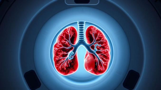
Researchers have developed a new MRI scanning method that provides real-time insights into lung function. This technique allows medical professionals to assess how effectively air moves through the lungs, offering valuable information for patients with asthma, COPD, and lung transplant recipients. The method uses perfluoropropane, a safe, inhalable gas, to pinpoint areas of effective and poor ventilation in the lungs.
See how TrueNAS offers real-time support for healthcare data managers.
** Main Story**
Okay, so, let’s talk about how Magnetic Resonance Imaging, or MRI, is shaking things up in respiratory care. It’s really quite fascinating, actually. We’re seeing a real shift in how we diagnose and treat lung diseases, and MRI is right at the forefront. This non-invasive imaging tech is giving us seriously detailed insights into lung function. Think earlier diagnoses, more targeted treatments – it’s a game changer for a bunch of respiratory conditions.
Why MRI is a Big Deal
Traditionally, you know, we’ve relied on spirometry. It measures how much air you can breathe in and out, and how fast. It’s useful, sure, but not exactly the most sensitive tool, especially when you’re trying to catch problems early. Plus, spirometry doesn’t tell you where the issue is in the lungs. Then there’s X-rays and CT scans. Great for seeing structure, but they expose patients to radiation. Not ideal for repeat monitoring, right?
MRI? Well, it’s different. It uses magnetic fields and radio waves to create high-resolution images of soft tissues, and importantly, no radiation. What’s really cool is the recent progress with inhaled gases, like perfluoropropane. These allow doctors to watch airflow and lung function in real time! Imagine seeing how oxygen is actually distributed throughout the lungs. That’s crucial for things like asthma and COPD. Honestly, it’s pretty impressive.
Catching Problems Sooner, Treating Them Better
Because MRI gives such a clear picture, we can spot lung problems way earlier, sometimes even before a patient notices symptoms. And early detection, it’s key. It means we can intervene sooner, slow down disease progression, and improve patient outcomes. That’s the goal, isn’t it?
Plus, and this is important, MRI helps us pinpoint exactly where the lung dysfunction is happening. This means, you guessed it, more personalized treatment. I remember reading a case study – or was it something my professor said, anyway – about a patient with asthma. They scanned him while he used his inhaler. The doctors could literally see how the medication affected airflow in different parts of his lungs. This allowed them to adjust the medication and dosage for maximum effectiveness and minimal side effects. Its personalized medicine in practice, and it really works.
More Than Just Asthma and COPD
It is worth noting that MRI is applicable far outside of just asthma and COPD. Think about it:
- Cystic Fibrosis: MRI can spot functional changes in the lungs of kids with CF even before standard lung function tests pick anything up.
- Interstitial Lung Disease: MRI is helping us understand and monitor the scarring in this disease.
- Pulmonary Vascular Disease: It’s improving diagnosis and management of problems with the blood vessels in the lungs.
- Post-COVID Lung Disease: Xenon MRI is being used to figure out the long-term lung effects of COVID-19. The results could be significant for our understanding of long term impacts.
- Clinical Trials: The sensitivity of MRI makes it an awesome tool for testing new lung medications.
The Road Ahead: AI and Beyond
These advances in MRI are changing how we do things now and paving the way for even more exciting developments. Think about artificial intelligence, AI, analyzing complex MRI data to find subtle patterns that could indicate early disease or predict when things might get worse. Imagine it, AI can flag things, that we, as humans might miss. Combine that with AI-powered wearable spirometry, and you’ve got a future where lung conditions are managed in a much more personalized, accessible, and effective way. I, for one, am pretty excited to see where all this goes.


The use of perfluoropropane as an inhalable contrast agent is intriguing. Could you elaborate on its safety profile compared to other contrast agents used in medical imaging, particularly regarding potential long-term effects on lung tissue or systemic exposure?
That’s a great question regarding the safety profile! While perfluoropropane is generally considered safe due to its inert nature and rapid elimination from the body, long-term studies are always essential. It’s an area of ongoing research as we strive for the best possible patient outcomes in medical imaging. It is certainly something the medical community are working towards, to improve healthcare outcomes. It really is an intriguing area.
Editor: MedTechNews.Uk
Thank you to our Sponsor Esdebe
So, MRI’s giving us real-time lung movies now? Finally, a chance to see my lungs dancing after that questionable gas station sushi. Seriously though, non-invasive and radiation-free is a huge win, especially for repeat monitoring. Anyone else wondering if we can get this tech hooked up to VR for a truly immersive respiratory experience?