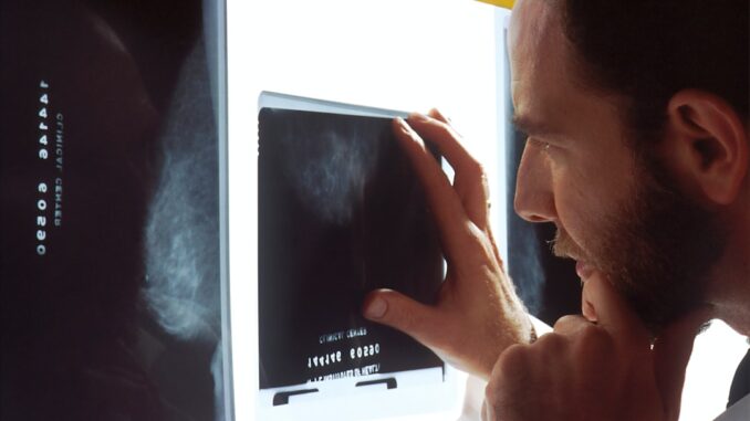
Shifting the Diagnostic Paradigm: AI Unlocks Fatty Liver Detection from Routine Chest X-Rays
Imagine a world where one of the most prevalent yet silent diseases, fatty liver disease, could be caught early, not through expensive, specialized scans, but from a simple, routine chest X-ray. Sounds like something out of a futuristic medical drama, doesn’t it? Well, thanks to pioneering work by researchers at Osaka Metropolitan University, this isn’t science fiction anymore. They’ve developed an AI model that truly could revolutionize how we screen for and manage this escalating global health crisis.
For far too long, diagnosing hepatic steatosis—the formal term for fatty liver—has felt a bit like chasing shadows. You see, the disease often presents with no symptoms in its early stages, subtly, progressively causing damage. When symptoms do appear, they’re often vague, things like fatigue or a dull ache in the upper right abdomen. By then, the condition might have already progressed to more serious stages. And the tools we’ve relied on? They’re effective, yes, but they carry their own set of challenges.
Healthcare data growth can be overwhelming scale effortlessly with TrueNAS by Esdebe.
Traditionally, a definitive diagnosis has hinged on advanced imaging techniques. Think ultrasound, CT scans, or even the gold standard, MRI. These modalities, while providing detailed insights into liver fat content, aren’t exactly perfect for widespread screening. They’re costly, often require specialized equipment and personnel, and let’s be honest, getting an appointment can be a real logistical hurdle. Plus, CT scans involve a dose of ionizing radiation, which you really want to limit, especially for screening purposes. This all creates a significant barrier to early detection, particularly in resource-constrained environments or for populations at higher risk who might need frequent monitoring.
The Silent Epidemic and Its Stakes
Before we dive deeper into this AI marvel, it’s vital to grasp the sheer scale of the problem we’re up against. Fatty liver disease, predominantly non-alcoholic fatty liver disease (NAFLD), affects an astonishing approximately 25% of the global population. That’s one in four people, a truly staggering figure, and its prevalence continues to climb, fueled by rising rates of obesity, type 2 diabetes, and metabolic syndrome. This isn’t just a minor health concern; it’s a silent epidemic with profound implications for public health worldwide.
Why should we be so concerned about a little extra fat on the liver? Because ‘little’ can quickly become ‘life-threatening’. If left unchecked, NAFLD can progress. It can morph into non-alcoholic steatohepatitis, or NASH, where inflammation and liver cell damage occur. From there, it’s a slippery slope to fibrosis, which is scarring of the liver tissue. And eventually, this scarring can lead to cirrhosis, an irreversible condition where the liver is severely damaged and unable to function properly. Cirrhosis, my friends, significantly increases the risk of liver failure, necessitating a transplant, and tragically, hepatocellular carcinoma, or liver cancer. So, early detection isn’t just a good idea; it’s absolutely crucial for preventing severe outcomes and saving lives.
A New Lens: Chest X-Rays and AI Synergy
Enter the game-changer from Osaka. The brilliance of this research lies in its simplicity and accessibility. Chest X-rays are, well, everywhere. They’re routinely performed for a myriad of reasons, from pre-surgical checks to pneumonia diagnoses, and they’re relatively inexpensive. Moreover, they involve minimal radiation exposure compared to CT, making them an ideal candidate for widespread, opportunistic screening. You’re already getting one, why not glean more vital health information from it?
The team built a deep learning AI model, training it on a massive dataset: 6,599 standard chest X-ray images, pulled from 4,414 distinct patients. To teach the AI what ‘fatty liver’ looks like on an X-ray—a subtle visual signature, to be sure—they correlated these images with controlled attenuation parameter (CAP) scores. For those unfamiliar, CAP is a non-invasive measurement derived from transient elastography (often part of a FibroScan), providing a quantitative assessment of liver fat content. It’s essentially a proxy for how much fat is actually in the liver, giving the AI a reliable ground truth to learn from. This thoughtful approach to data labeling is what often separates truly impactful AI from mere academic exercises.
And the results? Frankly, they’re impressive. The AI model achieved an area under the receiver operating characteristic curve (AUC) ranging from 0.82 to 0.83. If you’re not knee-deep in statistics, let me tell you, an AUC value approaching 1.0 signifies exceptional diagnostic accuracy. An AUC in the low 0.80s is indicative of high confidence and reliability in detecting hepatic steatosis, placing it firmly in the realm of clinically useful tools. What this means for us is that the model can effectively distinguish between individuals with and without fatty liver disease, identifying those at risk with remarkable precision. This isn’t just a slight improvement; it’s a significant leap forward in leveraging existing infrastructure for a critical public health challenge.
The Wider Canvas: AI’s Growing Footprint in Medical Imaging
It’s important to frame Osaka’s achievement within the broader narrative of AI’s burgeoning role in medical imaging. The concept of AI assisting with, or even leading, diagnostic interpretation isn’t novel; it’s been a rapidly evolving field. We’ve seen significant strides in using AI to assess various liver conditions across different imaging modalities.
Just consider the evidence: A meta-analysis of 13 studies, for instance, powerfully demonstrated how AI significantly improved the diagnosis of NAFLD, NASH, and liver fibrosis. It’s a testament to the technology’s pattern-recognition capabilities, far exceeding what the human eye, even a highly trained one, can consistently achieve across vast datasets. Similarly, a comprehensive systematic review and meta-analysis published in BMC Medical Imaging highlighted that image-based AI models exhibited high diagnostic performance for fatty liver disease, boasting pooled sensitivity of 92% and specificity of 94%. Think about that for a moment: detecting 92% of cases, and correctly identifying 94% of healthy individuals. Those are numbers that clinicians dream of.
These prior successes, largely using more specialized scans like CT and MRI, laid the groundwork. What makes the Osaka study particularly exciting is its pivot to chest X-rays. You’ve heard of AI systems interpreting mammograms for breast cancer, or detecting early signs of lung nodules from chest CTs, haven’t you? This research extends that promise to a truly ubiquitous imaging platform, making the benefits of AI-enhanced diagnostics far more broadly accessible.
Breaking Down Barriers: Accessibility, Cost, and Global Reach
Now, let’s talk about the practical implications. The integration of this AI-driven approach goes far beyond just providing a diagnosis; it’s about fundamentally re-shaping healthcare delivery. By leveraging the already pervasive infrastructure of chest X-rays, healthcare systems can potentially implement widespread, population-level screening programs without the need for investing in additional, costly specialized equipment. Imagine the immediate impact on hospital budgets, on waiting lists for more advanced scans, and ultimately, on patient pathways.
This could be an absolute game-changer, especially in resource-limited settings. Think about rural clinics in developing nations, or even underserved urban communities, where access to sophisticated imaging technologies like MRI or transient elastography is severely restricted, if available at all. A simple chest X-ray machine is often one of the first pieces of diagnostic equipment established in such areas. Suddenly, with this AI, that common machine transforms into a potent screening tool for a chronic disease that often goes undiagnosed for too long, leading to catastrophic health outcomes. We’re talking about democratizing early detection, bringing advanced diagnostic capabilities to places that desperately need them.
And it’s not just about geography or economic factors. Consider the sheer convenience for patients. Who wouldn’t prefer a quick, low-cost X-ray during a routine check-up to a more involved, perhaps uncomfortable, specialized scan? This ease of access can significantly boost screening rates and patient compliance, helping us cast a wider net to identify at-risk individuals earlier than ever before. It’s not just about clinical efficiency, it’s about better patient engagement, too. Couldn’t you agree?
Navigating the Road Ahead: Limitations and Future Horizons
That said, it’s essential for us, as informed professionals, to look at this development with a balanced perspective. Like any groundbreaking research, this study from Osaka Metropolitan University isn’t without its limitations. The research was conducted retrospectively, meaning the AI model was trained and tested on existing, pre-collected data. While this is a common and necessary first step, it carries inherent risks.
For one, retrospective studies can be prone to selection bias. The data might not perfectly represent the diversity of the general population or real-world clinical scenarios. The AI’s performance, therefore, may vary when applied to different patient demographics, various X-ray machine manufacturers, or even different imaging protocols. Consider how subtle differences in patient positioning, exposure settings, or even the technician’s technique could influence an X-ray image; a real-world validation would need to account for all of that variation. Therefore, further prospective studies are absolutely necessary. These would involve testing the AI model in new, unseen patient populations, ideally in multiple clinical settings, across different regions of the world, to truly confirm the model’s generalizability, robustness, and real-world effectiveness. We’d want to see how it performs in a busy emergency room versus a routine outpatient clinic, for example.
Beyond validating performance, there are other considerations. Regulatory hurdles, like obtaining FDA clearance in the US or CE Mark in Europe, are significant. These processes involve rigorous testing and validation to ensure the AI’s safety, efficacy, and reliability in a clinical environment. While companies like Nanox are making strides, having recently received FDA clearance for their HealthFLD, an AI-based software for assessing fatty liver, it highlights the stringent path from research to widespread clinical adoption. Then there’s the integration into existing electronic medical records (EMR) systems, ensuring seamless workflow for radiologists and clinicians. And what about potential algorithmic bias? We must ensure these models are trained on diverse datasets to avoid perpetuating health disparities.
A Glimpse into the Future of Diagnostics
In conclusion, the application of AI to detect fatty liver disease through standard chest X-rays represents a truly significant advancement in medical diagnostics. It offers a non-invasive, incredibly cost-effective, and remarkably accessible method for early detection that could genuinely transform the global management of liver diseases. Imagine the public health impact, the countless lives that could be positively affected by earlier interventions and vastly improved patient outcomes. I’m telling you, this isn’t just about a new piece of software; it’s about fundamentally democratizing access to critical health insights. It underscores the incredible potential of artificial intelligence, not as a replacement for human medical expertise, but as a powerful augmentation tool, enabling us to see more, understand more, and ultimately, do more for our patients. The future of diagnostic medicine is undoubtedly one where human intuition and AI’s analytical power work hand-in-hand, pushing the boundaries of what’s possible, and honestly, that’s a future I’m incredibly excited to be a part of.


Considering the current reliance on CAP scores for AI training, how might the variability in CAP measurements across different devices or patient populations affect the AI model’s accuracy and generalizability?
That’s a fantastic point! The variability in CAP scores across different devices and populations is definitely a crucial factor. Standardizing CAP measurements and ensuring diverse datasets for training are essential steps to improve the AI model’s generalizability and accuracy across broader clinical settings. Thanks for bringing up this important aspect!
Editor: MedTechNews.Uk
Thank you to our Sponsor Esdebe
This is a promising development, particularly regarding accessibility in resource-limited settings. Could this approach be adapted to identify other indicators of metabolic health, potentially expanding the utility of routine chest X-rays even further?
That’s a fantastic question! Absolutely, exploring the potential to identify other metabolic health indicators from chest X-rays is an exciting avenue. Imagine using a routine X-ray to screen for early signs of heart disease or diabetes! Further research in this direction could really maximize the value of this common imaging technique.
Editor: MedTechNews.Uk
Thank you to our Sponsor Esdebe