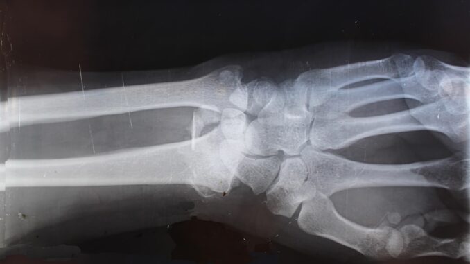
Unlocking the Unseen: How AI is Reshaping Osteoporosis Detection from Everyday X-rays
Imagine a silent thief, tirelessly working within your bones, subtly eroding their strength until, without warning, a simple stumble leads to a life-altering fracture. That’s osteoporosis, isn’t it? A pervasive, debilitating condition that too often goes unnoticed, its consequences only truly manifesting once irreversible damage has occurred. But what if we could catch this thief much earlier, perhaps even before it truly starts its destructive work? This isn’t science fiction anymore, not with the groundbreaking strides artificial intelligence is making in medical diagnostics.
Recently, in a truly compelling piece published in the Journal of Orthopaedic Research, an international team of researchers pulled back the curtain on an AI system. And what a system it is! It’s capable of estimating bone mineral density (BMD) directly from your run-of-the-mill, standard X-ray images. This isn’t just a neat trick; it’s an innovative approach poised, I’d say, to fundamentally revolutionize how we detect and manage osteoporosis, a condition known for weakening bones and, consequently, dramatically increasing fracture risk.
The Elusive Adversary: Why Osteoporosis Remains Undetected
Here’s the rub: osteoporosis often remains an invisible threat until a fracture shatters its secrecy. You see, the current gold standard for BMD measurement, dual-energy X-ray absorptiometry (DXA) scans, well, they’re not exactly ubiquitous. Their accessibility is often limited, and the cost can be quite prohibitive, especially in less developed regions or even in many rural parts of otherwise affluent countries. Think about it: a significant portion of the global population simply doesn’t have easy access to these specialized machines. It’s a real barrier, preventing millions of individuals worldwide from knowing they even have the condition.
This lack of widespread screening means countless people walk around unaware of their thinning bones, vulnerable to preventable fractures that lead to untold suffering, compromised quality of life, and, frankly, staggering healthcare costs. We’re talking about millions upon millions of dollars annually, much of which could be mitigated through earlier detection and intervention. It’s a systemic issue, and one that cries out for a more scalable, less resource-intensive solution.
The AI’s Keen Eye: Peering Through the Bone
Now, let’s talk about this remarkable AI system. The research team, harnessing the power of deep neural networks, crafted a tool designed to scrutinize routine X-ray images of the lumbar spine and femur. These aren’t just any old X-rays; these are the images clinicians order every single day for myriad reasons, from assessing back pain to checking on a twisted ankle. The brilliance lies in extracting profoundly insightful information from data we already collect.
They didn’t just guess, mind you. The system underwent rigorous training, learning from a massive dataset of 1,454 diverse X-ray images. It’s like teaching a child to recognize patterns, but on an infinitely more complex scale, feeding it example after example of both healthy and osteoporotic bones until it could discern the subtle, almost imperceptible nuances that signify bone density loss. And the results? They’re impressive, really quite striking:
- Sensitivity: For lumbar spine images, the system boasted an 86.4% sensitivity, meaning it was incredibly good at correctly identifying individuals who did have osteopenia (a precursor to osteoporosis). For femur images, it was similarly strong at 84.1%.
- Specificity: This is about avoiding false alarms. The system achieved an 80.4% specificity for lumbar spine images and 76.3% for femur images. This reflects its strong ability to correctly identify individuals who didn’t have osteopenia, saving them from unnecessary worry or follow-up procedures.
These numbers tell a compelling story. They suggest that this AI system can effectively pinpoint bone density loss, often even in patients who aren’t presenting with any overt symptoms. Think about the implications for proactive care! It’s like having an incredibly diligent, tireless second pair of eyes reviewing every X-ray that comes through, quietly flagging potential issues that might otherwise slip through the cracks of a busy clinical day.
Integrating Intelligence: A Seamless Shift in Clinical Practice
So, what does all this mean for the everyday practice of medicine? Well, integrating such an AI system into routine clinical workflows could be nothing short of a paradigm shift, facilitating earlier and far more widespread detection of osteoporosis. Picture this: a patient comes in for a routine chest X-ray for pneumonia, or perhaps an X-ray of their hip after a minor fall. That same image, processed by the AI in the background, could simultaneously provide a crucial, early warning about their bone health.
This approach aligns beautifully with the concept of ‘opportunistic screening.’ Instead of waiting for a patient to develop symptoms or for a physician to specifically order a DXA scan, we’re leveraging existing X-ray imaging infrastructure. Healthcare providers could identify at-risk individuals without the need for specialized equipment, without the added cost, and most importantly, without requiring additional patient visits. It’s about squeezing more value, more critical insight, out of procedures already being performed. This is not just efficient; it’s genuinely transformative patient care, making preventative medicine a truly seamless part of every interaction.
From Theory to Reality: AI’s Proven Impact
The potential of AI in osteoporosis detection isn’t just some theoretical pipe dream; it’s already showing tangible results in real-world applications. Take, for instance, a pivotal study highlighted in the Journal of the American College of Radiology. This research delved into the effectiveness of AI-enabled chest radiography analysis in uncovering osteoporosis. You see, the chest X-ray is one of the most common imaging procedures out there, and it often captures parts of the spine. The deep learning model used in that particular study successfully identified osteoporosis in over 75% of patients, aligning quite well with subsequent DXA findings. This wasn’t just a validation; it also facilitated a remarkable 10% higher detection rate compared to usual care. Just think of the impact of a 10% improvement when we’re talking about a condition that affects hundreds of millions globally!
This ability to opportunistically screen, essentially finding a hidden condition within an already ordered scan, is where the true power lies. It’s about turning every X-ray from a single-purpose diagnostic tool into a multi-purpose health scanner. And it’s a brilliant example of technology supporting, not replacing, the clinician, providing that extra layer of vigilance that human eyes, no matter how trained, might occasionally miss in the rush of a busy clinic.
The Long Road Ahead: Hurdles and Ethical Labyrinths
While the promise of this AI system shines brightly, it would be disingenuous to ignore the challenges that lie ahead. No groundbreaking technology arrives without its own set of complexities, does it?
One critical hurdle is ensuring the system’s accuracy across truly diverse populations and under varying imaging conditions. Our bones aren’t all built the same way, you know. There are inherent differences across ethnicities, age groups, and even lifestyle factors. Can the AI perform consistently whether it’s analyzing an X-ray from a state-of-the-art urban hospital or an older machine in a rural clinic? What about variations in patient positioning or the subtle differences in how different radiologists might acquire images? These are not minor points; they speak to the generalizability and robustness of the model. The training data must be comprehensive enough to account for this vast spectrum of human variability and imaging nuances.
Then there’s the monumental task of regulatory approval. We’re talking about a medical device, after all, something that could directly impact patient care and, yes, even liability. Navigating the stringent requirements of bodies like the FDA in the US or the CE marking in Europe demands rigorous validation, independent testing, and clear guidelines for deployment. It’s a meticulous, often lengthy process, but a crucial one for patient safety and physician trust.
Data privacy is another elephant in the room, isn’t it? As these AI systems ingest vast quantities of sensitive patient data, ensuring compliance with regulations like HIPAA and GDPR becomes paramount. How will data be anonymized? Where will it be stored? Who will have access? These are not just IT questions; they’re deeply ethical ones that speak to patient trust and the integrity of the healthcare system.
And let’s not forget the human element. Integrating AI tools into existing clinical workflows isn’t simply a matter of plugging in a new piece of software. It requires careful consideration of how clinicians—radiologists, primary care physicians, orthopedists—will interact with these tools. Will they trust the AI’s recommendations? Will they understand its limitations? There’s a vital need for training and education to ensure that these powerful AI ‘assists’ are used effectively and responsibly. We don’t want a situation where clinicians blindly follow AI advice, nor one where they ignore it entirely. It’s a delicate balance, this collaboration between human expertise and artificial intelligence.
Furthermore, the ‘black box’ problem, as some call it, can be a concern. While deep learning models are incredibly powerful, their decision-making processes can sometimes be opaque. For a clinician, understanding why the AI made a particular prediction can be just as important as the prediction itself, especially when dealing with life-altering diagnoses. Explainable AI (XAI) is an emerging field trying to address this, making AI’s reasoning more transparent. This will be key for widespread adoption in clinical settings.
Finally, we need to consider the economic implications. While opportunistic screening promises long-term cost savings by preventing costly fractures, the initial investment in developing, validating, and deploying these sophisticated AI systems isn’t trivial. The business case needs to be clearly articulated, demonstrating how this technology truly delivers value, not just a flashy new gadget.
The Horizon: A Future Shaped by AI in Bone Health
The development of AI systems capable of estimating BMD from routine X-rays represents a truly significant advancement in medical diagnostics. It’s a testament to human ingenuity, isn’t it, how we can repurpose existing tools to unlock entirely new diagnostic capabilities? By transforming standard imaging procedures—those everyday X-rays we barely give a second thought to—into powerful tools for osteoporosis detection, these technologies hold the potential to dramatically improve patient outcomes through earlier diagnosis and, crucially, earlier intervention.
Think about the ripples this could create. Imagine a future where routine check-ups passively screen for bone health, where at-risk individuals are identified years before they experience their first fracture, allowing for lifestyle changes, nutritional adjustments, and timely medication. This isn’t just about avoiding a broken hip; it’s about preserving independence, mobility, and the very quality of life for millions as they age. As research progresses, as these systems undergo further rigorous validation across broader, more diverse populations, I have no doubt that they will become integral components of osteoporosis screening programs worldwide.
Moreover, the future iterations of such AI models could move beyond just detection. Could they predict fracture risk with even greater precision? Perhaps monitor the efficacy of osteoporosis treatments over time, providing real-time feedback to clinicians? Or even tailor personalized treatment plans based on a patient’s unique bone structure and risk profile, integrating data points far too numerous for a human to process alone? The possibilities genuinely feel limitless.
Ultimately, this isn’t just about enhancing our diagnostic toolkit; it’s about proactively safeguarding public health. It’s about shifting the paradigm from reactive treatment to proactive prevention, using the best of human innovation to tackle one of our most pervasive silent diseases. The era of truly intelligent, integrated bone health management is truly dawning, and I, for one, can’t wait to see the profound impact it will have on countless lives.
References
- Moro, T., et al. (2025). Development of Artificial Intelligence‐Assisted Lumbar and Femoral BMD Estimation System Using Anteroposterior Lumbar X‐Ray Images. Journal of Orthopaedic Research. news-medical.net
- Bilbily, A., et al. (2024). Can AI Facilitate Improved Dual-Energy X-Ray Screening for Patients at High Risk of Osteoporosis? Journal of the American College of Radiology. diagnosticimaging.com


This is a fascinating exploration of AI’s potential. The discussion of “opportunistic screening” is particularly compelling; could this approach be expanded to detect other asymptomatic conditions during routine imaging, improving overall preventative care?
Thank you! That’s a great point. The potential for expanding opportunistic screening beyond osteoporosis is huge. Imagine detecting early signs of cardiovascular disease or even certain cancers from routine scans. It could truly revolutionize preventative care and improve health outcomes across the board. Let’s keep exploring these possibilities!
Editor: MedTechNews.Uk
Thank you to our Sponsor Esdebe
The discussion around AI’s potential to predict fracture risk and tailor treatment plans is exciting. Integrating diverse patient data points for personalized care could significantly improve outcomes beyond just early detection. How might we ensure equitable access to these advanced AI tools across different socioeconomic groups and healthcare systems?
Thanks for raising such a crucial point! The potential for personalized treatment plans based on diverse data is immense. Addressing equitable access is key; perhaps a tiered system based on healthcare infrastructure in the specific location could ensure broader availability. Thoughts?
Editor: MedTechNews.Uk
Thank you to our Sponsor Esdebe
The sensitivity and specificity results you shared are encouraging for integrating AI into routine X-ray analysis. Exploration of AI to proactively monitor treatment effectiveness and personalize treatment plans could be the next exciting advancement in osteoporosis management.
Thanks for highlighting the potential for personalized treatment plans! I agree that’s a key area for future development. Imagine AI tailoring medication dosages based on real-time bone density changes observed through routine X-rays. This level of precision could dramatically improve treatment outcomes and minimize side effects. What are your thoughts on the ethical considerations of AI-driven treatment adjustments?
Editor: MedTechNews.Uk
Thank you to our Sponsor Esdebe
AI quietly flagging potential issues is like having a diligent, tireless intern. But can we ensure the AI doesn’t develop a coffee addiction or start asking for a raise? I’m keen to explore how this tech supports, not replaces, clinicians, especially when the AI needs its own training!