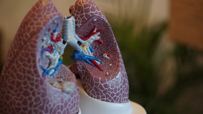
Robotic Bronchoscopy: A New Frontier in Lung Cancer Detection and Beyond
It’s no secret that lung cancer remains a formidable adversary in public health, often caught late when treatment options are limited. For far too long, diagnosing these elusive pulmonary lesions has presented a significant hurdle for clinicians, requiring invasive procedures that carry their own set of risks. But something truly transformative has been brewing in the medical field, a quiet revolution if you will. Robotic bronchoscopy, a cutting-edge technique, has emerged as a beacon of hope, fundamentally changing how we approach the detection and future treatment of lung cancer. It’s minimally invasive, incredibly precise, and frankly, a game-changer for patients.
The Lingering Challenge of Lung Lesions: Why Precision Matters
Before we dive into the marvels of robotics, it’s worth remembering the landscape we’ve been navigating. Think about traditional bronchoscopy: a crucial tool, absolutely, but one often wrestling with inherent limitations. For years, clinicians relied on what was essentially a flexible tube, threading it through the intricate, branching airways of the lungs. The challenge wasn’t just getting into the lung, but reaching those tiny, suspicious nodules nestled deep in the periphery, sometimes no bigger than a pea.
Secure patient data with ease. See how TrueNAS offers self-healing data protection.
Imagine trying to navigate a dense, three-dimensional maze with a rigid, somewhat unwieldy instrument. That’s what it felt like for many physicians. Peripheral lung nodules, those little troublemakers, are notoriously difficult to access. Their location, often in the very outermost reaches of the lungs, or nestled perilously close to vital structures like major blood vessels and the pleura, made biopsy a delicate, often imprecise dance. This frequently led to repeat procedures, higher complication rates, or worse, a diagnostic limbo where patients waited anxiously, uncertain about their prognosis. Moreover, the lack of real-time, comprehensive guidance often meant a ‘blind’ approach, hoping the instrument was truly within the lesion, not just brushing against its edge.
That’s where the frustration really set in, for both doctor and patient. You want to get that definitive diagnosis, you need to, but the tools simply weren’t always up to the task. It wasn’t uncommon to hear stories of a patient having multiple biopsies over months because the initial attempts simply couldn’t get enough, or the right, tissue. And that’s not just frustrating, it’s precious time ticking away.
Advancements in Robotic Bronchoscopy: A New Era of Access
So, what changed? Enter robotic-assisted bronchoscopy, a true paradigm shift. It’s not just an incremental improvement; it’s a quantum leap. This isn’t your science fiction movie robot stomping around, mind you, but a sophisticated system of ultrathin, highly maneuverable catheters, delicately guided by robotic arms. These aren’t just any arms either; they’re designed for dexterity that human hands simply can’t replicate within the tight confines of the bronchial tree. It allows clinicians to navigate the most intricate, tortuous bronchial pathways with unparalleled precision, reaching those previously inaccessible peripheral lung nodules. It’s like having a GPS system for the lungs, but with a micro-surgical tool at the end of it.
One of the real breakthroughs here is the integration of what’s called shape-sensing technology. Picture this: tiny optical fibers running through the catheter. As the catheter moves and bends, these fibers send back data, creating a real-time, three-dimensional map of the catheter’s exact position and shape within the lung. This isn’t static imaging; it’s dynamic, almost alive, giving the physician an immediate, intuitive sense of where they are and where they need to go. This level of granular control means physicians can steer the instrument through incredibly small and winding airways, past sharp turns, with a confidence that was previously unimaginable. This innovation has dramatically improved the diagnostic yield for peripheral lung lesions, with multiple studies, like one cited from pmc.ncbi.nlm.nih.gov, reporting success rates exceeding 80%. Imagine that: consistently hitting a target so tiny, so deep within the body. It’s phenomenal.
A Deeper Dive into the Technology: How It Works Its Magic
The robotic bronchoscopy platform typically consists of a console where the physician sits, essentially a cockpit with joysticks and a high-definition monitor displaying real-time images. This console is linked to a robotic arm system that holds and manipulates the flexible bronchoscope. But the magic really happens at the tip of the catheter, where the true innovation lies.
These catheters are remarkably thin, often less than 2mm in diameter, allowing them to traverse incredibly narrow bronchial branches. They incorporate technologies like electromagnetic navigation (EMN), which uses a low-frequency electromagnetic field to track the position and orientation of the catheter within the lung. This is integrated with pre-operative CT scans, essentially overlaying the real-time catheter position onto a 3D map of the patient’s unique lung anatomy. Think of it as a personalized topographical map, but for your bronchi.
Beyond EMN, some systems combine this with live fluoroscopy, providing a real-time X-ray view, and increasingly, with intra-operative cone-beam CT (CBCT). CBCT is particularly powerful because it allows for a quick, mini-CT scan during the procedure, confirming the catheter’s position relative to the lesion right then and there. This eliminates much of the guesswork. If the CBCT shows the catheter is slightly off, the physician can adjust it immediately, re-scan, and ensure perfect placement before taking the biopsy. This iterative process is what drives those impressive diagnostic yields, ensuring that the biopsy tool is precisely positioned within the lesion, not just adjacent to it. It’s truly a testament to engineering prowess.
Safety Profile and Patient Outcomes: A Breath of Fresh Air
Perhaps one of the most compelling aspects of robotic bronchoscopy, alongside its diagnostic prowess, is its remarkably favorable safety profile. Traditional bronchoscopy, while necessary, carried risks that could be quite serious. You’d worry about pneumothorax, that dreaded collapsed lung, or significant bleeding. These aren’t just minor inconveniences; they can mean extended hospital stays, additional procedures, and a longer, more painful recovery. I recall a case where a patient, already frail from their underlying condition, developed a pneumothorax after a standard biopsy. It set their treatment back weeks, and the emotional toll was palpable. It’s something you simply don’t want for anyone.
In stark contrast, robotic bronchoscopy has demonstrated a significantly lower incidence of these complications. For instance, a comprehensive study encompassing multiple centers, highlighted by medscape.com, found that device-related adverse events occurred in only 3.8% of patients. And let’s break that down further: pneumothorax requiring intervention was seen in just 2.8%, and bleeding events in a mere 0.9%. What’s truly remarkable, and comforting, is that no patients in that large study experienced severe respiratory failure. These numbers aren’t just statistics; they represent real people avoiding painful, life-threatening complications. It’s a huge step forward in patient safety, isn’t it?
This improved safety profile isn’t accidental. It’s a direct result of the robot’s enhanced precision and delicate touch. The controlled, measured movements of the robotic system minimize unnecessary tissue trauma. Less trauma means less chance of puncturing a delicate lung membrane or nicking a blood vessel. It’s like the difference between a blunt instrument and a surgeon’s scalpel, exquisitely controlled.
From a patient’s perspective, this translates into a much smoother, quicker recovery. The minimally invasive nature of the technique typically allows for same-day discharge, meaning patients can often be home for dinner, rather than spending days recuperating in a hospital bed. Think about the implications: reduced hospital stays, significant savings in healthcare costs, and perhaps most importantly, a faster return to daily activities and a better quality of life. Patients report less postoperative discomfort, less need for potent pain medication, and a general sense of ‘it wasn’t as bad as I thought it would be.’ It really is a testament to how far we’ve come in minimally invasive medicine.
Clinical Applications and Future Prospects: Beyond Just Biopsy
Robotic bronchoscopy isn’t just about improved diagnostic yield; it’s about unlocking new possibilities, especially for patients with lesions located in incredibly challenging areas. We’re talking about those nodules nestled precariously close to major blood vessels, where a traditional biopsy might carry an unacceptable risk of hemorrhage. Or lesions embedded within diseased lung tissue, perhaps scarred or emphysematous, which can make navigation a nightmare. The enhanced dexterity and fine control offered by robotic systems empower clinicians to perform biopsies in these high-risk zones with far greater confidence and safety. It’s like having microscopic hands that can maneuver where ours simply can’t, seeing with incredible clarity.
Moreover, the integration of real-time imaging technologies, like the mobile 3D imaging mentioned by mayoclinic.org, has further refined the accuracy of lesion targeting. This isn’t just a static image you’re looking at; it’s dynamic. Imagine seeing a live, three-dimensional representation of the lung, with your robotic instrument shown precisely within it. This ensures that the biopsy tools are positioned perfectly within the lesion, maximizing the chances of a definitive diagnosis on the first attempt.
But the exciting part? We’re only just scratching the surface of what robotic bronchoscopy can do. While diagnostic biopsy is its primary current application, the field is rapidly evolving towards therapeutic interventions. Imagine the potential to combine diagnostic and therapeutic procedures in a single session, under the same anesthesia. This isn’t just convenient; it’s profoundly impactful for patient care and outcomes. We could be talking about:
- Targeted Ablation: Using the same precise navigation to deliver therapies like radiofrequency ablation (RFA) or cryoablation directly to small, early-stage tumors. Instead of major surgery, you’re looking at a minimally invasive, localized treatment. Think about the reduced recovery time, the diminished risk.
- Drug Delivery: Precisely administering chemotherapy agents or novel immunotherapies directly into a tumor, potentially minimizing systemic side effects. It’s like a postal service for cancer drugs, delivering right to the doorstep.
- Fiducial Placement: Placing tiny markers, called fiducials, precisely within or around a tumor to guide highly accurate radiation therapy. This ensures the radiation beam hits its target perfectly, sparing healthy lung tissue.
- Airway Intervention: For non-cancerous conditions, the robot could assist in stent placement for airway strictures or even precisely deliver biologics for inflammatory lung diseases.
The Road Ahead: Innovation and Accessibility
Looking ahead, the landscape of robotic bronchoscopy continues to evolve at a breathtaking pace. Researchers are relentlessly working to refine these systems, making them even smaller, more flexible, and perhaps even incorporating haptic feedback, allowing the physician to ‘feel’ the tissue resistance through the controls. This would add another layer of intuition and safety.
The goal, of course, is to push diagnostic yields even higher, perhaps consistently into the 90%+ range, and to broaden the therapeutic toolkit available through this platform. Artificial intelligence is another fascinating frontier. AI algorithms could potentially assist with image analysis, predict optimal navigation pathways, and even help auto-segment lesions, further streamlining the procedure and enhancing accuracy. Could AI eventually ‘learn’ the nuances of navigating a specific patient’s unique lung anatomy? It’s a tantalizing thought.
However, we also need to address the practicalities. The widespread adoption of this technology hinges on several factors: the cost of the systems themselves, ensuring adequate training for pulmonologists and thoracic surgeons, and making sure this incredible technology is accessible to all who could benefit, not just those in major academic centers. It’s a powerful tool, but like any cutting-edge technology, scaling it up takes effort and investment.
In conclusion, robotic bronchoscopy represents not just a significant advancement, but a true revolution in the diagnostic approach to lung lesions. Its undeniable safety profile, remarkable precision, and minimally invasive nature offer substantial benefits over traditional methods, fundamentally improving patient experience and outcomes. This isn’t just about finding cancer earlier; it’s about doing so with less risk, less discomfort, and paving the way for more effective, patient-friendly diagnostic and therapeutic procedures in pulmonary medicine. We’re truly entering an exciting era, and frankly, I can’t wait to see what comes next.


Given the enhanced precision and safety profile, how might robotic bronchoscopy influence screening guidelines and the frequency of lung cancer screenings, especially for high-risk populations?
That’s a great question! With improved safety and precision, robotic bronchoscopy could definitely lead to more frequent screenings, especially for high-risk groups like smokers or those with a family history. Perhaps we’ll see guidelines evolve to incorporate this less invasive option for earlier detection and intervention, ultimately improving outcomes. What are your thoughts?
Editor: MedTechNews.Uk
Thank you to our Sponsor Esdebe
The integration of AI for image analysis and navigation is particularly exciting. Could this technology eventually enable more personalized approaches, tailoring the bronchoscopy procedure to an individual’s unique lung anatomy and lesion characteristics?
That’s a fantastic point! The potential for personalized approaches is huge. AI could analyze a patient’s unique lung structure and even predict lesion characteristics, enabling tailored navigation strategies and treatment plans. It could truly revolutionize how we approach lung cancer diagnosis and therapy. Thanks for sparking that thought!
Editor: MedTechNews.Uk
Thank you to our Sponsor Esdebe