Summary
MRI is revolutionizing the diagnosis and treatment of brachial plexus birth injuries in infants, offering earlier and more accurate assessments of nerve damage. This non-invasive imaging technique aids in determining the need for surgical intervention, reducing parental anxiety and potentially improving long-term outcomes. By providing a clearer picture of the injury, MRI empowers medical professionals to make informed decisions and tailor treatment plans for each child.
Secure patient data with ease. See how TrueNAS offers self-healing data protection.
Main Story
Pediatric care’s always moving forward, isn’t it? And right now, one area seeing some truly impressive advancements is how we diagnose and treat brachial plexus injuries in newborns. We’re talking about brachial plexus birth injury, or BPBI, which affects roughly one in every thousand babies. It’s not fun: it causes weakness or even paralysis in their upper extremities due to, sadly, trauma during childbirth.
Now, most of these little ones bounce back with minimal intervention, which is a relief. But, around 30% end up with lasting impairments. That’s where things get tricky. Figuring out which infants actually need surgery has been a real challenge. Traditionally, it’s meant months of just…watching, waiting, and a whole lot of anxiety for the parents. However, magnetic resonance imaging – MRI – is stepping up as a really powerful tool. It’s helping clear up the mystery around these injuries and guiding us toward the right treatment plans.
MRI: Seeing the Unseen
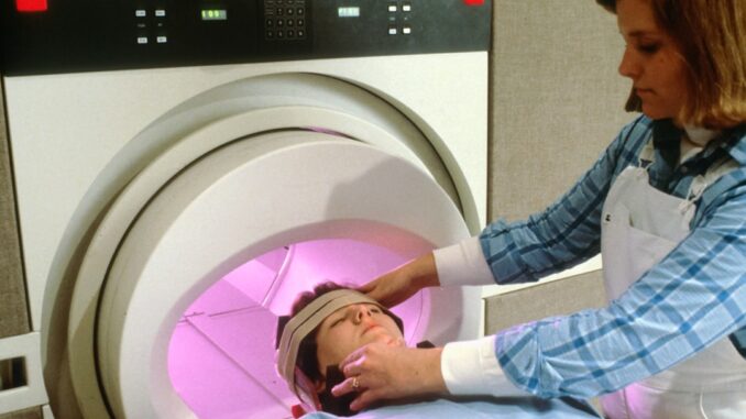
For years, diagnosing BPBI meant relying on physical exams and electrodiagnostic studies. And while they’re helpful, they often don’t give us the whole picture, especially in tiny infants. An MRI though? It’s a non-invasive way to actually see the brachial plexus – that crucial network of nerves controlling movement and sensation in the shoulder, arm, and hand.
Think of it like this: you’re trying to fix a broken wire, but you can’t see where the break is. An MRI is like a flashlight, showing you exactly where the damage lies.
Advanced MRI techniques, like high-resolution 3D-STIR SPACE sequences (try saying that three times fast!), allow us to really get into the details. We can assess the nerves and surrounding tissues, pinpointing the location and extent of the injury with amazing accuracy. In fact, studies show that MRI has a sensitivity and accuracy of around 88% in diagnosing brachial plexus injuries. That’s pretty darn good.
Early Diagnosis: A Game Changer
What’s so great about all this? Well, one of the biggest advantages of using MRI for BPBI is that it gives us early, accurate information about how severe the injury is. This is huge because early intervention can drastically improve long-term outcomes. If we can spot nerve root avulsions or ruptures early on, the MRI helps us figure out which babies are most likely to benefit from surgical repair.
That, in turn, reduces the need for prolonged observation and lets us intervene quickly, which can maximize the chances of restoring function. Plus – and this is something you can’t put a price on – early diagnosis eases the uncertainty and stress for families facing this challenging situation. I remember a case from a few years back. The parents were beside themselves, not knowing what to expect. Once we got the MRI results, even though it showed the injury was significant, they told me they felt a sense of relief just knowing what they were dealing with and having a plan.
Making It Easier on Families
Here’s another piece of good news: researchers have developed MRI protocols that don’t require sedation or contrast agents. That means the procedure’s safer and more comfortable for the infants. And that’s a big win. I mean, who wants to sedate a newborn if they don’t have to?
It’s especially important for families who might have to travel a distance to reach specialized brachial plexus centers. Non-sedated MRI means fewer visits and less disruption to their lives. And you know, travel costs add up, especially if it’s unexpected; the reduced visits can really ease some financial strain.
More Than Just Diagnosis
The thing is, MRI isn’t just about diagnosing BPBI. It’s also about guiding the treatment strategies. Think of it as a roadmap to recovery.
The NAPTIME study – it stands for Non-Anesthetized Plexus Technique for Infant MRI Evaluation – is a multicenter research effort that’s developed a scoring system. Based on the MRI findings, we can assess how extensive the BPBI is and predict whether surgery’s needed. It’s a standardized approach that helps clinicians make more objective, informed decisions about the best course of action for each child. And that? That’s huge.
The Road Ahead
Even though MRI is becoming more and more essential in managing BPBI, research is ongoing. Scientists are constantly working to improve the accuracy and predictive power of MRI findings. They’re exploring new techniques and developing better scoring systems. The hope is that this research will further demystify brachial plexus injuries and improve outcomes for the little ones affected.
Really, MRI is changing the game when it comes to BPBI care. By giving us a clearer view of the nerve damage, speeding up diagnosis, and guiding treatment decisions, it empowers us to provide the best possible care for these vulnerable infants. As technology keeps improving, MRI is poised to play an even bigger role in improving the lives of children with brachial plexus injuries. It’s an exciting time, and it makes me hopeful for what the future holds for these kids and their families.


High-resolution 3D-STIR SPACE sequences! Sounds like something straight out of a sci-fi movie, not medical imaging. I bet even the doctors need a cheat sheet to remember that one. Glad to hear it helps little ones though!
Haha, you’re right! “High-resolution 3D-STIR SPACE” does sound like something from Star Trek! It’s a mouthful, but the level of detail it gives us for diagnosing those tricky brachial plexus injuries in infants is invaluable. Early intervention can make such a difference. We are very glad the MRI helps little ones as well.
Editor: MedTechNews.Uk
Thank you to our Sponsor Esdebe
So, MRIs are now fortune tellers, predicting the need for surgery? Can it also predict if my toddler will *actually* eat the vegetables I sneak into their pasta sauce? Asking for a friend (who is me).
That’s a great question! While MRIs excel at visualizing nerve damage, predicting toddler food preferences might require a whole new level of AI. Perhaps a taste-bud scanner is next on the innovation list? Good luck with the pasta sauce!
Editor: MedTechNews.Uk
Thank you to our Sponsor Esdebe
The point about MRI reducing the need for sedation is significant. Could this also open doors to wider applications of MRI in pediatric care, particularly for conditions where sedation poses challenges?