Abstract
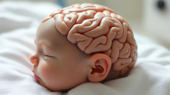
Encephalopathy encompasses a broad spectrum of neurological disorders characterized by altered brain function, leading to diverse clinical manifestations. This research report provides a comprehensive overview of encephalopathy, exploring its various classifications, underlying mechanisms, diagnostic approaches, and therapeutic strategies. While the discussion spans across different etiologies of encephalopathy, it maintains a particular focus on Hypoxic-Ischemic Encephalopathy (HIE), a significant cause of neonatal morbidity and mortality. Beyond the established treatment modalities such as therapeutic hypothermia, the report delves into emerging therapeutic avenues, preventative measures, and personalized treatment strategies, considering the influence of gestational age on treatment outcomes in HIE. We critically evaluate recent research advancements, discussing the potential of novel neuroprotective agents, regenerative medicine approaches, and strategies targeting inflammation and oxidative stress. Furthermore, the report addresses the challenges associated with encephalopathy management and highlights areas requiring further investigation to improve patient outcomes.
Many thanks to our sponsor Esdebe who helped us prepare this research report.
1. Introduction
Encephalopathy, derived from the Greek words ‘enkephalos’ (brain) and ‘pathos’ (suffering), is a general term denoting any diffuse disease of the brain that alters its function or structure. This alteration manifests clinically as changes in mental status, ranging from subtle cognitive deficits to profound coma. The etiology of encephalopathy is varied, including infectious agents, metabolic disorders, toxic exposures, traumatic brain injury, and hypoxic-ischemic events. The presentation and severity of encephalopathy depend on the underlying cause, the extent of brain involvement, and the patient’s age and pre-existing conditions.
Among the various causes of encephalopathy, Hypoxic-Ischemic Encephalopathy (HIE) stands out as a significant concern, particularly in the neonatal population. HIE results from a reduction in oxygen supply to the brain (hypoxia) coupled with insufficient blood flow (ischemia), leading to neuronal damage and subsequent neurological deficits. In neonates, HIE often occurs during the perinatal period due to complications such as placental abruption, umbilical cord compression, or difficult deliveries. The incidence of HIE is estimated to be 1-8 per 1000 live births, and it is a leading cause of cerebral palsy, cognitive impairment, and epilepsy in children. Despite advances in neonatal care, HIE remains a significant challenge, necessitating ongoing research into effective preventative and therapeutic strategies.
This research report aims to provide a comprehensive overview of encephalopathy, with a particular focus on HIE. We will explore the different classifications of encephalopathy, delve into the pathophysiology of HIE, critically evaluate current treatment options, and examine emerging research into preventative measures and alternative therapies. Furthermore, we will discuss the impact of gestational age on treatment effectiveness and explore personalized approaches to HIE management.
Many thanks to our sponsor Esdebe who helped us prepare this research report.
2. Classifications of Encephalopathy
Encephalopathy can be classified based on various criteria, including etiology, clinical presentation, and the reversibility of the condition. Understanding these classifications is crucial for accurate diagnosis and appropriate management.
2.1. Etiological Classification
Encephalopathy can be classified based on its underlying cause. Some common etiological categories include:
- Toxic-Metabolic Encephalopathy: This category encompasses encephalopathies resulting from exposure to toxins (e.g., alcohol, heavy metals, drugs) or metabolic disturbances (e.g., hepatic encephalopathy, uremic encephalopathy, hypoglycemia, hyperammonemia). These conditions often disrupt neuronal function through various mechanisms, such as altered neurotransmitter activity, mitochondrial dysfunction, or increased oxidative stress.
- Infectious Encephalopathy: Infections of the brain parenchyma (encephalitis) or meninges (meningitis) can lead to encephalopathy. Common causative agents include viruses (e.g., herpes simplex virus, West Nile virus), bacteria (e.g., Streptococcus pneumoniae, Neisseria meningitidis), and fungi (e.g., Cryptococcus neoformans). The inflammatory response triggered by the infection contributes to neuronal damage and dysfunction.
- Hypoxic-Ischemic Encephalopathy (HIE): As previously mentioned, HIE results from a reduction in oxygen supply and blood flow to the brain. This is particularly relevant in neonates but can also occur in adults following cardiac arrest, stroke, or near-drowning events. The pathophysiology of HIE involves a complex cascade of events, including energy failure, excitotoxicity, oxidative stress, and inflammation.
- Traumatic Encephalopathy: Traumatic brain injury (TBI) can cause acute or chronic encephalopathy. Acute encephalopathy following TBI is often due to direct neuronal damage, cerebral edema, and hemorrhage. Chronic Traumatic Encephalopathy (CTE) is a progressive neurodegenerative disease associated with repetitive head trauma, characterized by the accumulation of tau protein in the brain.
- Autoimmune Encephalopathy: This category includes encephalopathies caused by autoimmune processes that target the brain. Examples include autoimmune encephalitis associated with antibodies against neuronal surface antigens (e.g., NMDA receptor encephalitis, LGI1 encephalitis) and paraneoplastic neurological syndromes. The autoimmune response leads to neuronal dysfunction and inflammation.
- Genetic Encephalopathy: Certain genetic disorders can cause encephalopathy. These include metabolic disorders (e.g., phenylketonuria, maple syrup urine disease), mitochondrial disorders, and neurodegenerative diseases (e.g., Huntington’s disease, Alzheimer’s disease). The genetic mutations disrupt neuronal function through various mechanisms.
2.2. Clinical Classification
Encephalopathy can also be classified based on its clinical presentation. This classification is often based on the severity of symptoms and the presence of specific neurological findings.
- Acute Encephalopathy: This refers to a rapid onset of altered mental status, often accompanied by other neurological signs such as seizures, focal deficits, or movement disorders. Acute encephalopathy requires prompt evaluation to identify the underlying cause and initiate appropriate treatment.
- Subacute Encephalopathy: This refers to a more gradual onset of altered mental status, developing over days to weeks. Subacute encephalopathy can be caused by various conditions, including infections, autoimmune disorders, and metabolic disturbances.
- Chronic Encephalopathy: This refers to a long-standing and persistent alteration in mental status. Chronic encephalopathy can be caused by neurodegenerative diseases, chronic toxic exposures, or structural brain abnormalities.
- Static Encephalopathy: This describes a condition where brain damage has occurred and is not progressing. The neurological deficits remain relatively stable over time.
- Progressive Encephalopathy: This describes a condition where brain damage continues to worsen over time. This is often associated with neurodegenerative diseases or ongoing toxic exposures.
2.3. Reversibility Classification
Another important classification considers the reversibility of the encephalopathy. Some encephalopathies are reversible with prompt and appropriate treatment, while others may lead to permanent neurological deficits.
- Reversible Encephalopathy: This refers to encephalopathies that can be completely resolved with treatment. Examples include encephalopathies caused by drug overdoses, metabolic disturbances, or infections that respond well to therapy.
- Partially Reversible Encephalopathy: This refers to encephalopathies where some improvement in neurological function can be achieved with treatment, but some residual deficits may persist.
- Irreversible Encephalopathy: This refers to encephalopathies that result in permanent brain damage and neurological deficits. Examples include severe HIE, advanced neurodegenerative diseases, and extensive traumatic brain injury.
Many thanks to our sponsor Esdebe who helped us prepare this research report.
3. Hypoxic-Ischemic Encephalopathy (HIE): Focus on Neonatal Aspects
Hypoxic-ischemic encephalopathy (HIE) in neonates results from a period of oxygen deprivation and reduced blood flow to the brain around the time of birth. This can lead to significant brain damage and long-term neurological disabilities. Understanding the pathophysiology, diagnosis, and management of HIE is crucial for improving outcomes in affected infants.
3.1. Pathophysiology of HIE
The pathophysiology of HIE is complex and involves a cascade of events that lead to neuronal injury. The initial insult of hypoxia and ischemia triggers a series of cellular and molecular processes, including:
- Energy Failure: Oxygen deprivation disrupts mitochondrial function, leading to a decrease in ATP production. This energy failure impairs cellular processes, including ion homeostasis and neurotransmitter transport.
- Excitotoxicity: Energy failure leads to depolarization of neurons and the release of excessive amounts of glutamate, an excitatory neurotransmitter. Overstimulation of glutamate receptors, particularly NMDA receptors, results in an influx of calcium into neurons, leading to excitotoxicity and neuronal death.
- Oxidative Stress: Hypoxia-ischemia leads to the production of reactive oxygen species (ROS), which cause oxidative damage to cellular components, including lipids, proteins, and DNA. Oxidative stress contributes to neuronal injury and apoptosis.
- Inflammation: Hypoxia-ischemia activates inflammatory pathways, leading to the release of cytokines and chemokines. These inflammatory mediators contribute to neuronal damage and edema formation.
- Apoptosis: Programmed cell death (apoptosis) is a major mechanism of neuronal injury in HIE. Apoptosis is triggered by various factors, including excitotoxicity, oxidative stress, and inflammation.
3.2. Diagnosis of HIE
The diagnosis of HIE is based on a combination of clinical findings, laboratory tests, and neuroimaging studies. Clinical criteria include:
- History of perinatal asphyxia: Evidence of a hypoxic-ischemic event around the time of birth, such as placental abruption, umbilical cord compression, or difficult delivery.
- Apgar scores: Low Apgar scores at 5 and 10 minutes of age.
- Umbilical artery blood gas analysis: Acidosis (pH < 7.0) and base deficit.
- Clinical signs of encephalopathy: Altered level of consciousness, seizures, hypotonia, and abnormal reflexes.
Laboratory tests may include:
- Blood glucose: To rule out hypoglycemia.
- Electrolytes: To assess for electrolyte imbalances.
- Liver function tests: To evaluate liver function.
- Renal function tests: To evaluate renal function.
- Coagulation studies: To assess for bleeding disorders.
Neuroimaging studies, such as magnetic resonance imaging (MRI) and amplitude-integrated electroencephalography (aEEG), play a crucial role in confirming the diagnosis of HIE and assessing the extent of brain damage. MRI can identify specific patterns of injury, such as watershed infarcts, basal ganglia injury, and cortical injury. aEEG can monitor brain activity and detect seizures.
3.3 Current Treatment Options for HIE (Excluding Cooling Therapy)
While therapeutic hypothermia (cooling therapy) is the standard of care for HIE, it is not effective in all cases, and it may not be available in all settings. Therefore, other treatment options are essential for managing HIE.
- Supportive Care: Supportive care is crucial for all infants with HIE. This includes maintaining adequate oxygenation, ventilation, and blood pressure. Fluid and electrolyte management is also important to prevent complications such as cerebral edema.
- Seizure Management: Seizures are common in infants with HIE and can contribute to further brain damage. Antiepileptic medications, such as phenobarbital or levetiracetam, are used to control seizures.
- Management of Cerebral Edema: Cerebral edema can worsen brain damage in HIE. Strategies to manage cerebral edema include elevating the head of the bed, restricting fluid intake, and administering diuretics such as mannitol.
- Nutritional Support: Adequate nutrition is essential for brain recovery in HIE. Infants with HIE may require parenteral nutrition or enteral feeding through a nasogastric tube.
3.4. New Research into Preventative Measures and Alternative Treatments
Research into preventative measures and alternative treatments for HIE is ongoing. Some promising areas of investigation include:
- Antenatal Corticosteroids: Antenatal corticosteroids are commonly used to promote lung maturation in premature infants. Some studies suggest that antenatal corticosteroids may also have neuroprotective effects in infants at risk for HIE.
- Magnesium Sulfate: Magnesium sulfate has been shown to have neuroprotective effects in animal models of HIE. Some clinical trials have suggested that magnesium sulfate may reduce the risk of cerebral palsy in infants born prematurely or at risk for HIE.
- Erythropoietin (EPO): Erythropoietin is a hormone that stimulates red blood cell production. EPO has also been shown to have neuroprotective effects in animal models of HIE. Several clinical trials are investigating the potential of EPO to reduce brain injury in infants with HIE. A meta-analysis of studies showed that EPO combined with hypothermia did not improve outcome or reduce mortality rates for infants with HIE. Some did however, report a reduction in MRI detectable brain injury. [1]
- Xenon: Xenon is an inert gas that has neuroprotective properties. Studies have shown that xenon can reduce brain injury in animal models of HIE. Clinical trials are investigating the potential of xenon as an adjunct therapy to hypothermia in infants with HIE.
- Stem Cell Therapy: Stem cell therapy involves the transplantation of stem cells into the brain to promote tissue repair and regeneration. Stem cell therapy is a promising but still experimental treatment for HIE. The exact mechanisms of action of stem cell therapy in HIE are not fully understood, but it is believed that stem cells can release growth factors, reduce inflammation, and promote angiogenesis. It has been demonstrated in trials involving stem cell therapy that stem cells do not differentiate into neurons as initially theorised. [2]
Many thanks to our sponsor Esdebe who helped us prepare this research report.
4. Gestational Age and Treatment Effectiveness
Gestational age is a significant factor influencing the effectiveness of HIE treatment. Preterm infants are more vulnerable to brain injury from HIE than term infants. This is because their brains are still developing and are more susceptible to oxidative stress and inflammation.
- Therapeutic Hypothermia: Therapeutic hypothermia is most effective when initiated within 6 hours of birth. However, the optimal duration and target temperature of hypothermia may vary depending on gestational age. Some studies suggest that preterm infants may benefit from longer durations of hypothermia or lower target temperatures. This is an area of active research [3].
- Neuroprotective Agents: The effectiveness of neuroprotective agents, such as EPO and magnesium sulfate, may also vary depending on gestational age. Further research is needed to determine the optimal dosing and timing of these agents in preterm and term infants with HIE.
Many thanks to our sponsor Esdebe who helped us prepare this research report.
5. Future Directions and Challenges
Despite advances in the management of HIE, significant challenges remain. Future research should focus on:
- Developing more effective neuroprotective agents: Current neuroprotective agents have limited efficacy in preventing brain damage in HIE. More research is needed to identify novel therapeutic targets and develop more effective neuroprotective strategies.
- Personalizing treatment strategies: Treatment strategies for HIE should be tailored to the individual patient, considering factors such as gestational age, severity of HIE, and underlying comorbidities.
- Improving long-term outcomes: Long-term follow-up of infants with HIE is essential to monitor their neurodevelopmental outcomes and provide appropriate interventions. Research is needed to identify factors that predict long-term outcomes and develop strategies to improve outcomes.
- The Role of Biomarkers: Early identification of HIE is important to improve prognosis. Research into Biomarkers which can identify the condition early is still an ongoing and emerging field. Several biochemical markers such as hypoxanthine, lactate, S100B protein, neuron-specific enolase (NSE), creatine kinase-BB (CK-BB) have been identified, as well as miRNAs. [4] Developing tools to easily identify HIE will improve outcome in infants.
- Novel Therapeutic Targets: Emerging evidence suggests that targeting specific cellular and molecular pathways involved in the pathophysiology of HIE may hold promise for improving outcomes. For example, inhibiting inflammatory pathways, reducing oxidative stress, or promoting neuronal survival may be effective strategies.
- Regenerative Medicine Approaches: Regenerative medicine approaches, such as stem cell therapy and gene therapy, have the potential to repair damaged brain tissue and restore neurological function in infants with HIE. However, these approaches are still in the early stages of development, and further research is needed to evaluate their safety and efficacy.
Many thanks to our sponsor Esdebe who helped us prepare this research report.
6. Conclusion
Encephalopathy is a complex and heterogeneous group of disorders that can result from various etiologies. HIE, in particular, poses a significant challenge in neonates, leading to long-term neurological disabilities. While therapeutic hypothermia has improved outcomes in HIE, it is not universally effective, and other treatment options are needed. Emerging research into preventative measures and alternative treatments, such as antenatal corticosteroids, magnesium sulfate, EPO, and stem cell therapy, holds promise for improving outcomes in HIE. Future research should focus on developing more effective neuroprotective agents, personalizing treatment strategies, and improving long-term outcomes. Overcoming these challenges will require a multidisciplinary approach involving clinicians, researchers, and policymakers.
Many thanks to our sponsor Esdebe who helped us prepare this research report.
References
[1] Yang, S., Chen, Q., & Zhou, W. (2023). Erythropoietin as an adjuvant to hypothermia for hypoxic-ischemic encephalopathy: A meta-analysis and trial sequential analysis. Frontiers in Pediatrics, 11, 1125743. https://doi.org/10.3389/fped.2023.1125743
[2] Donegan, C., & Levy, M. L. (2020). Stem cell therapy for neonatal hypoxic-ischemic encephalopathy. Current Opinion in Pediatrics, 32(2), 217-223.
[3] Tagin, M. A., Woolcott, C. G., Vincer, M. J., Whyte, R. K., Stinson, D. A., Moore, A. M., … & Canadian Neonatal Network Investigators. (2003). Hypothermia for neonatal hypoxic ischemic encephalopathy: an updated systematic review and meta-analysis. Archives of Disease in Childhood-Fetal and Neonatal Edition, 88(5), F346-F356.
[4] Toledano-Alhadef, H., & González, A. S. (2018). Biomarkers in Hypoxic-Ischemic Encephalopathy. Frontiers in Neurology, 9, 876. https://doi.org/10.3389/fneur.2018.00876


Stem cell therapy…so, are we talking brain ‘re-boot camp’ where cells learn to adult properly? Maybe they get tiny diplomas and then start contributing to society (or, you know, just stop causing trouble in the brain neighborhood). Intriguing stuff!
That’s a fantastic analogy! “Brain re-boot camp” really captures the essence of the hope behind stem cell therapy. The idea of guiding these cells to ‘contribute to society’ in the brain is what researchers are striving for. It’s fascinating to consider the potential for targeted interventions to improve brain health and function. What are your thoughts on the ethical considerations surrounding this?
Editor: MedTechNews.Uk
Thank you to our Sponsor Esdebe