Unlocking Bone Health: How AI is Revolutionizing Osteoporosis Detection from Routine X-rays
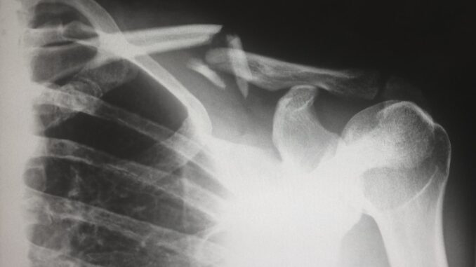
Imagine a world where the insidious creep of osteoporosis, that silent thief of bone density, no longer goes undetected until it’s too late. It’s a future that’s rapidly coming into focus, thanks to some truly groundbreaking work from researchers at the University of Tokyo. In a recent study, published in the Journal of Orthopaedic Research, they unveiled an artificial intelligence system capable of estimating bone mineral density (BMD) directly from routine X-ray images. It’s an innovation, truly, that just might reshape how we approach osteoporosis screening, leveraging those common X-rays of the lumbar spine and femur that we’re already taking for a myriad of other reasons. Isn’t that something? Talk about efficiency and impact.
This isn’t just about tweaking an existing process; it’s about potentially revolutionizing early detection and intervention strategies for a condition that affects millions globally. You see, the implications here are profound, promising to democratize access to crucial bone health insights, pushing us closer to a future where bone fractures, a devastating consequence of advanced osteoporosis, become far less common.
Osteoporosis: The Silent Epidemic and Its Cost
Let’s be candid for a moment about osteoporosis. It’s a disease often characterized as ‘silent’ for a reason. It gnaws away at skeletal strength, gradually, relentlessly, without obvious symptoms until a significant event, like a fracture, finally brings it to light. By then, quite a bit of damage has already occurred, hasn’t it? We’re talking about weakened bones and a tragically increased risk of fractures, often from seemingly minor falls. A cough, even, can sometimes be enough to break a rib in someone severely affected. Think about the impact: chronic pain, loss of mobility, decreased quality of life, and for too many, premature mortality. The statistics are stark, aren’t they? One in three women and one in five men over the age of 50 will experience an osteoporotic fracture in their lifetime.
Traditionally, assessing BMD, our gold standard for diagnosing osteoporosis and its precursor, osteopenia, involves a dual-energy X-ray absorptiometry, or DXA scan. Now, DXA is incredibly accurate, we can’t argue with that. It precisely measures bone density at key sites like the hip and spine, providing a T-score that tells us just how far a patient’s BMD deviates from that of a healthy young adult. It’s the benchmark, the trusted tool that guides our clinical decisions.
The Gold Standard: DXA and Its Limitations
But here’s the rub: while DXA is accurate, it isn’t always accessible, is it? You find clinics with long waiting lists, especially in areas with fewer specialized facilities. Cost can be a barrier for some, even if insurance often covers it for high-risk individuals. Then there’s the patient experience; it requires a dedicated appointment, often a referral, and the machine itself can be somewhat intimidating. For many, particularly those living in rural or underserved communities, getting to a DXA machine can be a logistical nightmare, not to mention the direct cost or co-pay. This inherent lack of widespread accessibility directly contributes to underdiagnosis and, predictably, delayed treatment. It’s a systemic challenge we’ve grappled with for years.
Consider this scenario: A busy GP sees hundreds of patients a week. They know which ones are at higher risk for osteoporosis – perhaps an older patient, a post-menopausal woman, someone with a history of certain medications or chronic conditions. They might order an X-ray for back pain or a suspected fracture. But to then order another scan, a DXA, requires a separate process, another appointment, often weeks later. It adds friction, doesn’t it? That friction is where patients fall through the cracks.
The Untapped Potential of Routine X-rays
Routine X-rays, on the other hand, are ubiquitous. Almost every clinic, every hospital, has an X-ray machine. They’re performed millions of times a day globally for everything from assessing lung infections to diagnosing sprains and fractures. The sheer volume and availability make them incredibly appealing. Yet, until now, we haven’t been able to effectively harness them for precise BMD estimation. Why? Because the subtle changes in bone density that signify osteopenia or osteoporosis aren’t easily discernible to the human eye on a standard plain film X-ray. A radiologist can spot obvious bone loss in severe cases, yes, but for early detection, when intervention truly makes the most difference, it’s simply too challenging, too subjective.
This is where the magic of AI steps in, offering a bridge between the vast accessibility of X-rays and the diagnostic precision we need for early detection. It’s like giving X-rays a new superpower, isn’t it?
The AI Breakthrough: Unpacking the Tokyo Study
Steering this innovative charge, as you know, was the research team led by Dr. Toru Moro from the University of Tokyo. Their work builds upon years of research, not just their own, but also the collective efforts of the scientific community striving to unlock diagnostic potential from seemingly commonplace medical images. Dr. Moro’s team really focused on the lumbar spine and femur – critical areas because these are common sites for osteoporotic fractures and, conveniently, are routinely X-rayed for a host of other musculoskeletal complaints.
Crafting the Intelligent Eye: Dataset and Training
Developing an AI model capable of this feat isn’t a trivial undertaking. It requires meticulous attention to data, a cornerstone of any robust AI system. The team compiled a dataset comprising 1,454 X-ray images. Now, that number might not sound enormous in the age of ‘big data,’ but for medical imaging, particularly when each image needs careful annotation and correlation with a precise DXA measurement, it represents a substantial effort. Each X-ray image of the lumbar spine or proximal femur was paired with a corresponding DXA scan from the same patient, providing the AI with its ‘ground truth’ – the actual BMD values it needed to learn from.
Think of it this way: the AI, likely a type of deep learning model, probably a convolutional neural network (CNN), was fed these pairs of images and data points. It learned to identify subtle, complex patterns and features within the X-ray images that correlate with varying levels of bone density. These aren’t patterns that are obvious to us, but the AI, through millions of iterative adjustments to its internal parameters, finds these incredibly nuanced visual cues. It’s a bit like teaching a child to recognize different types of trees, but instead of ‘oak’ or ‘maple,’ it’s learning ‘dense bone’ or ‘porous bone’ from shadows and textures that are almost imperceptible to the human eye. The model was trained to estimate the T-score and even classify patients into categories like ‘normal,’ ‘osteopenia,’ or ‘osteoporosis,’ based solely on those X-rays.
When they put the system to the test, it truly demonstrated impressive performance. The system showed high sensitivity and specificity in identifying patients with osteopenia and osteoporosis. What does that mean for us in practical terms? High sensitivity means it’s very good at correctly identifying those with the condition, minimizing false negatives – we won’t miss many patients who need attention. High specificity means it’s also very good at correctly identifying those without the condition, reducing false positives – we won’t unnecessarily alarm or over-refer patients who are healthy. This dual strength indicates its strong potential as a reliable screening tool.
Why This Study Stands Out
It’s important to place this study in context. The idea of estimating BMD from X-rays isn’t entirely new, but previous efforts often faced hurdles. For instance, some research, like that by Zheng et al. (2021) or Wang et al. (2022), focused on hip X-rays or even chest X-rays. While valuable, this particular study by Dr. Moro’s team zeroes in on lumbar spine and femur X-rays – imaging types that are already incredibly common for a myriad of reasons beyond bone health. This isn’t about using a novel image type; it’s about extracting more value from images we’re already capturing. Gu et al. (2022, 2023) also explored advanced techniques like GANs and decomposition to estimate BMD, pushing the boundaries of what’s possible. However, the Tokyo team’s direct focus on widely performed anatomical regions for typical X-rays, and their clear demonstration of high sensitivity and specificity, presents a remarkably pragmatic and immediately implementable solution.
This isn’t just an academic exercise; it’s about harnessing existing infrastructure. It’s about saying, ‘Hey, we’ve got all these X-rays being taken anyway. Can we glean more diagnostic information from them, particularly for a silent killer like osteoporosis?’ And the answer, increasingly, seems to be a resounding ‘yes.’
Transforming Clinical Workflows: A Glimpse into the Future
Now, let’s talk about the real-world impact. Integrating this AI system into routine clinical workflows could be nothing short of transformative. Imagine a scenario: a patient comes in for a lower back issue, and a lumbar spine X-ray is performed. While the clinician is reviewing the image for disc space narrowing or vertebral alignment, the AI, running quietly in the background, simultaneously analyzes the image for BMD. If it detects signs of osteopenia or osteoporosis, it could immediately flag the patient’s record, perhaps even suggesting a follow-up DXA scan or a consultation with an endocrinologist.
Integration and Accessibility: Breaking Down Barriers
This seamless integration means no extra appointments, no new imaging modalities, and crucially, no additional radiation exposure beyond the initial diagnostic X-ray. It’s a ‘two birds, one stone’ approach that’s incredibly appealing. And think about the sheer accessibility it provides. Because it leverages existing X-ray equipment, this AI system could be deployed virtually anywhere an X-ray machine is present – from major urban hospitals to small community clinics, even remote healthcare facilities. This radically levels the playing field, making advanced screening capabilities available in places where DXA machines are a distant dream.
The Tangible Benefits: From Early Detection to Proactive Care
The benefits cascade from there. Earlier detection of osteoporosis means timely interventions become possible. This isn’t just about prescribing medication, though that’s often a crucial part of the puzzle. It means clinicians can initiate vital conversations about lifestyle modifications – dietary changes to increase calcium and vitamin D intake, weight-bearing exercise routines, fall prevention strategies. It means guiding patients towards proactive care rather than reactive treatment after a debilitating fracture has already occurred. Wouldn’t you agree that preventing a fracture is always preferable to treating one?
From an economic standpoint, its ability to analyze standard X-rays makes it a remarkably cost-effective option for widespread screening. We’re not talking about purchasing millions of dollars in new DXA equipment; we’re talking about a software overlay, a smart algorithm, that plugs into existing digital imaging systems (PACS). This significantly reduces the financial barrier to entry for healthcare providers and, by extension, for patients. Moreover, by catching osteoporosis earlier, we can hopefully mitigate the immense societal costs associated with fracture care, rehabilitation, and long-term disability. The economic burden of osteoporotic fractures is staggering, so any tool that can reduce that load is worth its weight in gold.
Navigating the New Landscape: Workflow Adjustments and Ethical Considerations
Of course, like any new technology, this isn’t simply a plug-and-play solution. Its introduction will necessitate careful consideration of clinical workflows. How will radiologists and other clinicians interpret these AI-generated insights? Will it create an ‘alert fatigue’ if too many flags are raised? We’ll need clear protocols for confirmatory DXA scans and patient communication. It’s about augmenting human expertise, not replacing it. The AI highlights, the human confirms and acts.
Then there are the ethical considerations. Data privacy, for one, is paramount. How is patient data secured as it flows through these AI systems? And what about algorithmic bias? Could the AI perform differently across various demographic groups or ethnicities if the training data wasn’t sufficiently diverse? These are critical questions that must be addressed rigorously as the technology matures and moves towards widespread adoption. Ensuring fairness and equity in AI deployment is, I think, a moral imperative, wouldn’t you say?
The Road Ahead: Validation, Adoption, and Beyond
While the initial results from Dr. Moro’s team are undeniably promising, let’s remember this is the cutting edge. Like any pioneering medical technology, further validation studies are absolutely necessary. This isn’t a ‘nice-to-have’; it’s a ‘must-have.’ We need to confirm the system’s accuracy across vastly diverse populations – different age groups, ethnicities, body types, and indeed, across patients with various underlying health conditions that might influence bone health or X-ray image quality. Does it perform equally well on images from different X-ray machine manufacturers or with varying exposure settings? These are practical questions that demand robust, large-scale clinical trials.
The Imperative of Further Research
Longitudinal studies will also be crucial. We need to see if this early detection truly translates into improved patient outcomes, such as reduced fracture rates, over time. It’s one thing to accurately detect a condition; it’s another to demonstrate that detecting it earlier makes a tangible difference in a patient’s life. Think about it: does flagging a patient at 55 with early osteopenia via AI-powered X-ray lead to interventions that prevent a hip fracture at 70? That’s the ultimate goal, isn’t it?
Regulatory Hurdles and Market Readiness
Beyond scientific validation, there are significant regulatory hurdles to clear. Medical AI systems, especially those involved in diagnosis, face stringent approval processes from bodies like the FDA in the US or the EMA in Europe. This ensures patient safety and efficacy. These processes are lengthy and demanding, requiring extensive documentation, rigorous testing, and transparent reporting. It’s a necessary step, but one that adds time and cost to market readiness. Then there’s the challenge of adoption within the healthcare system. Clinicians, understandably, want to trust the tools they use. Building that trust requires not just strong data, but also intuitive interfaces, robust technical support, and clear guidelines for use. It’s a complex ecosystem of technology, human behavior, and institutional inertia.
The Broader Picture: AI’s Role in Preventive Medicine
Nevertheless, this advancement represents a significant stride towards leveraging AI to improve bone health diagnostics and, consequently, patient outcomes. It’s part of a broader trend where AI is increasingly being deployed to unlock hidden insights from existing medical data, moving us towards a more proactive, preventive model of healthcare. Imagine other conditions that could be screened this way – early signs of heart disease from chest X-rays, perhaps, or subtle indicators of metabolic disorders. The possibilities are genuinely exhilarating.
Conclusion: Building a Stronger Future, One Bone at a Time
What we’re witnessing here isn’t just an incremental improvement; it’s a paradigm shift. By imbuing routine X-rays with the intelligence to assess bone mineral density, Dr. Moro’s team isn’t just solving a diagnostic challenge; they’re opening a door to more equitable and accessible healthcare for millions at risk of osteoporosis. It’s a powerful demonstration of how AI, when applied thoughtfully and rigorously, can truly empower clinicians and patients alike.
We’re not quite at the finish line, but the path forward looks clearer than ever. This intelligent system promises to convert widely available, often underutilized, X-ray data into actionable insights, making bone health a more transparent and manageable aspect of preventive care. It’s an exciting time to be in healthcare, isn’t it? As we continue to refine and validate these remarkable AI tools, we’re not just building better algorithms; we’re building a stronger, healthier future, one bone at a time.
References
- Moro, T., et al. (2025). Development of Artificial Intelligence‐Assisted Lumbar and Femoral BMD Estimation System Using Anteroposterior Lumbar X‐Ray Images. Journal of Orthopaedic Research. (news-medical.net)
- Zheng, K., et al. (2021). Semi-Supervised Learning for Bone Mineral Density Estimation in Hip X-ray Images. arXiv preprint. (arxiv.org)
- Wang, F., et al. (2022). Lumbar Bone Mineral Density Estimation from Chest X-ray Images: Anatomy-aware Attentive Multi-ROI Modeling. arXiv preprint. (arxiv.org)
- Gu, Y., et al. (2022). BMD-GAN: Bone Mineral Density Estimation Using X-ray Image Decomposition into Projections of Bone-Segmented Quantitative Computed Tomography Using Hierarchical Learning. arXiv preprint. (arxiv.org)
- Gu, Y., et al. (2023). Bone Mineral Density Estimation from a Plain X-ray Image by Learning Decomposition into Projections of Bone-Segmented Computed Tomography. Medical Image Analysis. (arxiv.org)


The potential for integrating this AI into standard clinical workflows is particularly compelling. Could this technology also be adapted to analyze dental X-rays, offering an even earlier and more accessible screening opportunity for osteoporosis?
That’s a fantastic point! Using dental X-rays could indeed offer an even earlier and more accessible screening for osteoporosis. It would be great if future research explores this avenue. The lower radiation exposure and frequency of dental X-rays could make this a valuable screening tool.
Editor: MedTechNews.Uk
Thank you to our Sponsor Esdebe
AI with X-ray vision for bones? Talk about a glow-up! If AI can spot osteoporosis from routine X-rays, what’s next? Maybe it can also diagnose that embarrassing sock tan.